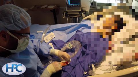
- Як виконувати внутрішньокісткову катетеризацію, вручну і з електросверлом.
- Як виконувати катетеризацію периферичної вени
- Як виконувати катетеризацію периферичної вени під ультразвуковим контролем
- Як виконувати катетеризацію променевої вени
- Як виконувати катетеризацію променевої артерії за допомогою ультразвукового контролю
- Як виконувати взяття проби венозної крові
Ресурси за темою
(See also Vascular Access.)
The radial artery is the most frequent site of arterial catheterization.
When ultrasonographic equipment and trained personnel are available, ultrasonographic guidance may be helpful in cannulating nonpalpable arteries (eg, due to obesity or a small artery) and increases the success rate of radial artery cannulation. This topic will focus on the use of ultrasonography to guide arterial cannulation. The actual procedure for radial artery cannulation is the same as when ultrasonography is not used and is described in detail in How To Do Radial Artery Cannulation.
Indications for US-Guided Radial Artery Cannulation
Difficulty in localizing the radial artery by palpation
Contraindications to US-Guided Radial Artery Cannulation
Absolute contraindications
None
Relative contraindications
There are some relative contraindications for radial artery cannulation, but once an appropriate site is identified, there are no contraindications to use of ultrasonography other than
Untrained or inexperienced ultrasound operator
Complications of US-Guided Radial Artery Cannulation
None
There are a number of complications of radial artery cannulation, but these are unrelated to use of ultrasonography.
Equipment for US-Guided Radial Artery Cannulation
In addition to standard equipment needed to cannulate the radial artery, operators will need the following:
Ultrasound machine with high frequency (eg, 5 to 10 MHz or higher), linear array probe (transducer)
Sterile, water-based lubricant, single-use packet (preferred over multi-use bottle of ultrasound gel)
Sterile probe cover, to ensheathe the probe and probe cable, sterile rubber bands (alternatively, the probe may be placed within a sterile glove and the cord wrapped within a sterile drape)
Additional Considerations for US-Guided Radial Artery Cannulation
Arterial catheterization is done under universal (barrier) precautions and sterile conditions.
The short-axis (transverse, cross-sectional) ultrasound view is easy to obtain and is the best view for identifying veins and arteries and their orientation to each other. However, the transverse view also shows the needle in cross-section (hyperechoic [white] dot), and the needle tip can be distinguished only by the appearance and disappearance of the white dot as the imaging plane traverses past the needle tip.
The long-axis (longitudinal, in-plane) ultrasound view is technically more difficult to obtain (must keep probe, vein, and needle in one plane), but the entire needle (including the tip) is imaged continuously, which ensures accurate intraluminal placement.
The narrowness of the radial artery increases the difficulty of obtaining the longitudinal view.
Relevant Anatomy for US-Guided Radial Artery Cannulation
The radial artery lies close to the skin over the ventrolateral distal wrist, just medial to the radial styloid process and lateral to the flexor carpi radialis tendon. The artery runs deeper in the more proximal wrist and the forearm.
Positioning for US-Guided Radial Artery Cannulation
Position the patient comfortably reclined or supine.
Rest the patient's forearm supinated and with the wrist extended on the bed or on a bedside table; support may be useful under the wrist.
Stand or sit at the side of the bed so that your nondominant hand is proximal on the arm with the artery to be cannulated; this allows the natural movement of your dominant hand to insert the catheter in a proximal direction.
Position the ultrasound console in the vicinity of the ipsilateral shoulder, so that you can see both it and the cannulation site without having to turn your head.
Step-by-Step Description of US-Guided Radial Artery Cannulation
The procedure for preparing the site and inserting and securing the radial artery catheter is the same as when ultrasonographic guidance is not used and is not described in full here.
Підготуйте ультразвуковий пристрій і визначіть променеву артерію
Check that the ultrasound machine is configured and functioning correctly: Set the machine to 2-D mode or B mode. Ensure that the screen image correlates with the spatial orientation of the probe as you are holding and moving it. The side-mark on the probe corresponds to a marker dot/symbol on the ultrasound screen. Adjust the screen settings and probe position if needed to attain an accurate left-right orientation.
Do a preliminary ultrasound inspection (nonsterile) of the area to determine whether the site is suitable for cannulation. Use a transverse (cross-sectional, short-axis) view, and set the depth until the radius is just visualized at the far field of the screen (depth markers are displayed on the side of the screen). Adjust the gain on the console so that the blood vessels are anechoic (appear black on the ultrasound screen) and the surrounding tissues are gray. Arteries are generally smaller, thick-walled, and round (rather than thin-walled and ovoid) and are less easily compressed (by pressing the probe against the skin) than veins. After identifying the radial artery, adjust the depth so that it is positioned in the middle third of the screen.
Use color Doppler mode to identify a patent lumen and spectral Doppler mode to identify pulsatile blood flow in the artery.
Some clinicians do the Allen test to determine whether there is sufficient collateral flow through the ulnar artery to perfuse the hand if the catheter occludes the radial artery. While the patient makes a tight fist, digitally compress both the ulnar and radial arteries. While continuing arterial compression, have the patient open the fist and spread the fingers, which should display a blanched palm and fingers. Then, release ulnar artery compression while maintaining radial artery compression. If the hand and fingers on the radial side reperfuse within 5 to 10 seconds, collateral circulation is considered adequate. Alternatively, ascertain the presence of ulnar artery flow by palpation or Doppler evaluation.
Supinate the forearm and tape both the hand and the mid-forearm to a dorsally placed arm board, with a gauze roll placed under the wrist to maintain moderate wrist extension.
Підготуйте обладнання і стерильне поле
Assemble the arterial pressure–monitoring equipment: Place the IV saline bag within the pressure bag (unpressurized), connect the arterial pressure tubing to the saline bag, and squeeze residual air from the bag into the line. Hang the bag, pinch the drip chamber to fill it halfway with fluid, and run solution through the tubing to flush the air out. Connect (plug in) the pressure transducer to the pressure monitor. Situate the transducer at the level of the heart (ie, lateral to the intersection of the mid-axillary line and 4th intercostal space). Open the transducer to air, set the transducer signal to zero on the monitor, and then close the transducer to air. Ensure all air is flushed from the tubing. Remove all vent caps and replace with sealed caps at all the ports. Then pressurize the bag to 300 mm Hg. Throughout the process, maintain sterility of all connecting points of the tubing.
Place sterile equipment on sterilely covered equipment trays.
Dress in sterile garb and use barrier protection.
Test the equipment: Rotate the catheter about the needle and slide the guidewire into and out of the needle to verify smooth motion. Push and pull syringe plungers to establish free motion and expel air from the syringes.
Draw the local anesthetic into a 3-mL syringe with 25-gauge needle attached.
Swab the ventral wrist area broadly with antiseptic solution (eg, chlorhexidine/alcohol).
Allow the antiseptic solution to dry for at least 1 minute.
Place sterile towels and large drapes about the site (large drapes are for maintaining sterility of the ultrasound probe and cord).
Покладіть стерильне покривало зверху ультразвукового датчика
Direct your assistant (nonsterile) to apply ultrasound gel (nonsterile) and then hold the probe, with the probe footprint pointing up, just outside the sterile field.
Insert your gloved dominant hand into the sterile probe cover.
Drape the sterile probe cover over the probe, by first grasping the probe with your dominant (covered) hand and then using your nondominant hand to unroll the sterile cover down over the probe and probe cable. Do not touch the uncovered cord or allow it to touch the sterile field as you unroll the cover.
Pull the cover tightly over the probe footprint to eliminate all air bubbles.
Wrap sterile rubber bands around the probe to secure the cover in place. The probe may now rest on the sterile drapes.
Виконайте анестезію місця катетеризації
Apply sterile ultrasound gel to the covered probe footprint.
Ultrasound guidance (transverse view) may be used for the lidocaine injection to avoid a vascular puncture.
Inject 1 to 2 mL of anesthetic into the skin and subcutaneously along the anticipated needle-insertion path.
Maintain gentle negative pressure on the syringe plunger as you advance the needle, to identify intravascular placement and prevent an intravascular injection.
Уведіть голку в променеву артерію за допомогою ультразвукового контролю
Using your nondominant hand, place the probe on the skin, always proximal to the anticipated needle-insertion point.
Always maintain ultrasound visualization of the needle tip during insertion.
Obtain an optimal cross-sectional (transverse) image of the radial artery in the distal forearm, and position the artery in the center of the screen.
Hold the cannulation device between the thumb and forefinger of your dominant hand.
Orient the needle bevel facing up.
Initially, slightly slide the ultrasound probe distally from the target artery entrance site to guide (lead) the needle from the skin insertion site to the target artery entrance proximally. Aim the needle at about a 30- to 45-degree angle into the skin and toward the midpoint of the probe. Hold the needle stationary once skin is punctured. Fan the ultrasound probe to identify the needle tip. The needle is hyperechoic (appears as a white dot on the ultrasound screen in the transverse view).
Advance the cannulation device. You may prefer to maintain the transverse view throughout the cannulation. Slightly tilt the probe fore-and-aft as you advance the needle, to continually reidentify the needle tip (disappearing/reappearing white dot as you tilt [fan] the probe). Or, you may prefer to switch to the longitudinal (long-axis) view (shown in the video) to see the needle and artery lengthwise. Turn the probe 90 degrees and maintain full longitudinal (in-plane) images of both the needle (including the tip) and the artery.
Advance the cannulation device into the artery. As the cannulation device approaches the artery, decrease the angle of insertion so the needle tip will enter with as much control as possible and on a more shallow angle to the artery. You should see the needle first indenting the superficial arterial wall and then popping through the wall to enter the lumen. A simultaneous flash of bright red, pulsatile blood in the reservoir or barrel of the device confirms intra-arterial placement.
Keep the cannulation device stationary in this spot.
Follow standard procedures for inserting and threading the catheter, ensuring intraarterial placement, securing it, dressing the site, and beginning pressure monitoring.
Warnings and Common Errors for US-Guided Radial Artery Cannulation
Once the needle punctures the skin, it is no longer helpful to inspect the wrist. Instead, look at the ultrasound screen and move the probe to look for the needle tip.
During cardiopulmonary arrest or other conditions of low blood pressure and hypoxia, arterial blood may be dark and not pulsatile and may be mistaken for venous blood.
Tips and Tricks for US-Guided Radial Artery Cannulation
It is prudent to confirm placement by imaging the length of the catheter inside the radial artery prior to suturing the catheter in place.
If an assistant is not available, cover the ultrasound control panel with a clear sterile sheath to enable operation of the machine during the procedure.


