- Як виконувати внутрішньокісткову катетеризацію, вручну і з електросверлом.
- Як виконувати катетеризацію периферичної вени
- Як виконувати катетеризацію периферичної вени під ультразвуковим контролем
- Як виконувати катетеризацію променевої вени
- Як виконувати катетеризацію променевої артерії за допомогою ультразвукового контролю
- Як виконувати взяття проби венозної крові
Ресурси за темою
The radial artery is the most frequent site used for arterial catheterization.
Ultrasound guidance, when equipment and trained personnel are available, is helpful in cannulating nonpalpable arteries (eg, due to obesity or a small artery).
(See also Vascular Access and How To Do Radial Artery Cannulation, Ultrasound-Guided.)
Indications for Radial Artery Cannulation
In critically ill, unstable patients, especially those with refractory shock and respiratory failure, or in patients undergoing complex surgery with fluid shifts or blood loss:
Continuous blood pressure measurement
Repeated arterial blood gas measurements
Repeated blood sampling for laboratory tests
Contraindications to Radial Artery Cannulation
Absolute contraindications
An artery that is neither palpable nor detectable by ultrasonography (never try to cannulate a site simply because the artery is expected to be there)
Unsuitable artery (eg, dialysis fistula at same limb, thrombosed or inaccessible artery, generalized limb ischemia)
Inadequate collateral blood flow from ulnar artery circulation
Full-thickness burns
Local infection at the insertion site
Relative contraindications
Coagulopathy (including therapeutic anticoagulation*) or recent/pending thrombolysis: The radial artery is the preferred arterial cannulation site in these situations; prolonged pressure (eg, 10 minutes or more) may be needed to stop bleeding/hematoma at the site.
Local anatomic distortion (traumatic or congenital), or gross obesity
History of prior surgery in the area
Distal extremity ischemia/gangrene
* Therapeutic anticoagulation (eg, for atrial fibrillation) increases the risk of bleeding with radial artery cannulation, but this must be balanced against the increased risk of thrombosis (eg, stroke) if anticoagulation is reversed. Discuss any contemplated reversal with the clinician managing the patient's anticoagulation and then with the patient.
Complications of Radial Artery Cannulation
Complications include
Hematoma
Infection
Damage to the artery
Thrombosis (due to the catheter itself)
Nerve damage
Catheter misplacement
To reduce the risk of catheter-related infections, radial artery catheters should remain in place no more than 7 days and their transparent, occlusive dressings left undisturbed. Catheters should be removed as soon as they are no longer needed or if there are any signs of infection.
Rare complications include
Distal ischemia and necrosis
Pseudoaneurysm
Arteriovenous fistula
Cholesterol plaque (or atheroma); air, guidewire, or catheter embolism
Ischemia of the hand occurs rarely, due to thrombosis or embolism, intimal dissection, or arterial spasm. Collateral blood flow from the ulnar artery usually prevents significant ischemia. The risk of arterial thrombosis is higher in small arteries (explaining the greater incidence in women) and with increased duration of catheterization. The incidence of thrombosis and distal ischemia is much lower for femoral arterial catheterization. Occluded arteries nearly always recanalize after catheter removal.
Equipment for Radial Artery Cannulation
Sterile procedure, barrier protection
Antiseptic solution (eg, chlorhexidine, povidone-iodine)
Sterile drapes, towels
Sterile head caps, masks, gowns, gloves
Face shields
Radial artery cannulation
Arm board, gauze roll, and tape
Local anesthetic (eg, 1% lidocaine without epinephrine, 25-gauge needle, 3-mL syringe)
Sterile gauze (eg, 10 cm × 10 cm squares)
3-mL and 5-mL syringes
Cannulation device (eg, integrated catheter and guidewire device; separate needle, guidewire and catheter; or peripheral venous angiocatheter [catheter-over-needle], 20 or 22 gauge)
Blood pressure monitor; IV saline bag (500 mL), pressure bag, and hanger; integrated arterial pressure line or individual components (ie, pressure transducer, arterial line tubing [noncompliant pressure tubing], 3-way stopcocks)
Nonabsorbable suture (eg, 3-0 or 4-0 nylon or silk)
Chlorhexidine patch, transparent occlusive dressing
An assistant or two is helpful.
Additional Considerations for Radial Artery Cannulation
Arterial catheterization is done under universal (barrier) precautions and sterile conditions.
A failed attempt to cannulate the radial artery may be followed by attempts more proximally, but only if arterial spasm does not occur and the radial pulse remains palpable. If the pulse is lost, further cannulation attempts of the artery—as well as of the ipsilateral ulnar artery—are prohibited.
If the radial arteries cannot be cannulated, alternative arterial sites include brachial or dorsalis pedis arteries peripherally, or femoral or axillary arteries centrally.
Relevant Anatomy for Radial Artery Cannulation
The radial artery lies close to the skin over the ventrolateral distal wrist, just medial to the radial head and lateral to the flexor carpi radialis tendon.
Positioning for Radial Artery Cannulation
Position the patient comfortably reclined or supine.
Rest the patient's forearm supinated and with the wrist extended on the bed or a bedside table; support may be useful under the wrist.
Stand or sit at the side of the bed so that your nondominant hand is proximal on the arm with the artery to be cannulated; this allows the natural movement of your dominant hand to insert the catheter in a proximal direction.
Step-by-Step Description of Radial Artery Cannulation
Palpate the radial artery in detail with the tip of the index finger of your nondominant hand. Systematically palpate, release, and shift slightly over the artery, to precisely discern the center axis of the artery (the area of the strongest pulse).
Some clinicians do the Allen test to determine whether there is sufficient collateral flow through the ulnar artery to perfuse the hand if the catheter occludes the radial artery. While the patient makes a tight fist, digitally compress both the ulnar and radial arteries. While continuing arterial compression, have the patient open the fist and spread the fingers, which should display a blanched palm and fingers. Then, release ulnar artery compression while maintaining radial artery compression. If the hand and fingers on the radial side reperfuse within 5 to 10 seconds, collateral circulation is considered adequate. Alternatively, just ascertain the presence of ulnar artery flow by palpation or Doppler evaluation.
Supinate the forearm and tape both the hand and the mid-forearm to a dorsally placed arm board, with a gauze roll placed under the wrist to maintain moderate wrist extension.
Підготуйте обладнання і стерильне поле
Assemble the arterial pressure–monitoring equipment: Place the IV saline bag within the pressure bag (unpressurized), connect the arterial pressure tubing to the saline bag, and squeeze residual air from the bag into the line. Hang the bag, pinch the drip chamber to fill it halfway with fluid, and run solution through the tubing to flush the air out. Connect (plug in) the pressure transducer to the pressure monitor. Situate the transducer at the level of the heart (ie, lateral to the intersection of the mid-axillary line and 4th intercostal space). Open the transducer to air, set the transducer signal to zero on the monitor, and then close the transducer to air. Ensure all air is flushed from the tubing. Remove all vent caps and replace with sealed caps at all the ports. Then pressurize the bag to 300 mm Hg. Throughout the process, maintain sterility of all connecting points of the tubing.
Place sterile equipment on sterilely covered equipment trays.
Dress in sterile garb and use barrier protection.
Test the cannulation equipment: Rotate the catheter about the needle and slide the guidewire into and out of the needle to verify smooth motion. Push and pull syringe plungers to establish free motion and expel air from the syringes.
Draw the local anesthetic into a 3-mL syringe with a 25-gauge needle attached.
Swab the ventral wrist area broadly with antiseptic solution (eg, chlorhexidine/alcohol).
Allow the antiseptic solution to dry for at least 1 minute.
Place sterile towels and drapes about the site.
Виконайте анестезію місця катетеризації
Inject 1 to 2 mL of anesthetic into the skin and subcutaneously along the anticipated needle-insertion path. Do not make a skin bleb so big that it obscures palpation of the radial artery.
Maintain gentle negative pressure on the syringe plunger as you advance the needle, to identify intravascular placement and prevent an intravascular injection.
Уведіть голку в артерію
Relocate the radial artery in the wrist as described previously, using your nondominant hand, and continue palpation to guide needle insertion into the artery.
Using your dominant hand, hold the cannulation device between your thumb and forefinger. Orient the needle bevel facing up.
Insert the cannulation device with the needle bevel facing up directly over the midline of the radial pulse at least 1 cm proximal to the radial head and advance it proximally (cephalad) at about a 30- to 45-degree angle into the skin, to intersect the artery.
Steadily advance the cannulation device until a flash of bright red blood appears in the reservoir or barrel of the device, which indicates that the needle tip has entered the arterial lumen.
Hold the device motionless in this spot.
If no blood flash appears after inserting a catheter-over-wire device 1 to 2 cm, slowly and gradually withdraw the device. If it had initially passed completely through the artery, a blood flash may now appear as the needle tip is withdrawn, passing back into the lumen. If a flash still does not appear, withdraw the device almost to the skin surface, change direction, and try again to advance it into the artery.
If no blood flash appears after inserting an angiocatheter 1 to 2 cm, hold the catheter steady and slowly withdraw the needle from it. A blood flash may appear if the needle tip alone had pierced the deep arterial wall. If a flash does not appear, keep withdrawing the needle until it is removed, and then slowly withdraw the catheter. If a flash appears, stop withdrawing and try to advance the catheter into the artery (some operators insert a guidewire before readvancing the catheter, to facilitate the catheter's passage into the arterial lumen).
If rapid local swelling occurs, blood is extravasating. Terminate the procedure: Remove the needle and use gauze pads to hold external pressure on the area for 10 minutes or more, to help limit bleeding and hematoma.
Оцініть повернення крові
Place a gauze square under the cannulation device at the insertion site.
Observe the reservoir or barrel of the device to verify pulsatile blood flow. If needed, slightly advance or withdraw the device until the pulsatile flow is evident, which confirms intra-arterial placement.
Continuously hold the cannulation device motionless at this spot.
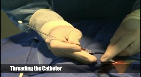
Просувайте артеріальний катетер
Catheter-over-wire technique
Thread the guidewire through the needle and into the artery. Do not force the wire; it should slide smoothly.
If the guidewire meets resistance, it may have passed into or through the arterial wall. Remove the catheter-over-wire device as a unit, use gauze pads for 10 minutes to apply pressure to the area (to help prevent bleeding and hematoma), and start over at a new insertion site with a new catheter-over-wire device.
Securely hold the needle hub and slide the catheter, using a twisting motion, over the needle and guidewire and into the artery.
Angiocatheter
The insertion method is essentially the same as starting an IV in a peripheral vein.
Further decrease the angle of insertion and advance the angiocatheter an additional 2 mm to ensure that the catheter tip has entered the lumen. This step is done because the needle tip slightly precedes the catheter tip.
Securely hold the needle hub and slide the catheter over the needle and into the artery; it should slide smoothly.
If the catheter meets resistance, slowly withdraw the needle followed by the catheter, stopping immediately and trying to re-advance the catheter if blood flow resumes. If the catheter cannot be inserted, withdraw it and start over. Never withdraw the catheter back over the needle or reinsert the needle back into the catheter (doing so may shear off the end of the catheter within the patient). Likewise, never withdraw the guidewire through the needle. Use gauze pads for 10 minutes to apply external pressure to the area.
Sometimes the catheter cannot be advanced although it is in the lumen; try to advance the catheter while flushing it with fluid from a syringe.
Під'єднайте артеріальну в/в систему
Attach the pressure tubing (which has been pre-flushed with saline) to the catheter hub and verify an arterial pressure waveform on the monitor screen.
Накладіть пов'язку на місце інфузії
Use gauze to wipe all blood and fluid from the site, being careful not to disturb the catheter.
Suture the catheter in place at the insertion site. To avoid skin necrosis, tie air loops in the skin and then tie the suture tails to the catheter hub.
Apply a transparent occlusive dressing. Chlorhexidine-impregnated discs at the insertion point are commonly placed before the dressing.
Loop the arterial tubing and tape it to the skin away from the insertion site, to help prevent accidental traction on the tubing from dislodging the catheter.
Write the date and time of cannulation on the dressing.
Warnings and Common Errors for Radial Artery Cannulation
Not carefully determining the exact center of the pulse makes it more likely to miss the artery.
Sliding your finger from side to side as you attempt to locate the point of maximum pulsation may give an inaccurate result; lift and reposition your finger laterally to each new location.
If the artery is not entered when the needle has reached an appropriate depth, do not try to reposition the needle by moving the tip to one side or another; this can damage tissue. Instead, withdraw the needle almost to the skin surface before changing the angle and direction of insertion.
Never inject drugs into an arterial line.
During cardiopulmonary arrest or other conditions of low blood pressure and hypoxia, arterial blood may be dark and not pulsatile and may be mistaken for venous blood.
Tips and Tricks for Radial Artery Cannulation
Avoid pushing too hard on the artery while palpating the pulse during needle insertion; doing so may compress the artery and make it harder to cannulate.
Some arterial lines are sensitive to wrist position; be alert to changes in the pressure and/or waveform with patient movement. The wrist may need to be immobilized in the most satisfactory position.


