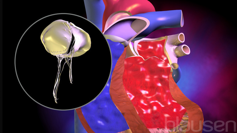
Mitral stenosis is a narrowing of the mitral valve opening that blocks (obstructs) blood flow from the left atrium to the left ventricle.
Mitral stenosis usually results from rheumatic fever, but infants can be born with the condition.
Mitral stenosis does not cause symptoms unless it is severe.
Doctors make the diagnosis after hearing a characteristic heart murmur through a stethoscope placed over the heart, and they use echocardiography to make a more detailed diagnosis.
Treatment begins with use of diuretics and beta-blockers or calcium channel blockers.
The valve can be stretched open with a catheter, but occasionally the valve needs to be replaced, requiring open-heart surgery.
(See also Overview of Heart Valve Disorders and the video The Heart.)
The mitral valve is in the opening between the left atrium and the left ventricle. The mitral valve opens to allow blood from the left atrium to fill the left ventricle and closes as the left ventricle contracts to prevent blood from flowing back into the left atrium as the ventricle pumps blood into the aorta. If a disorder causes the valve flaps to become thick and stiff, the valve opening is narrowed. Sometimes the stiffened valve also fails to close completely, and mitral regurgitation develops.
In mitral stenosis, blood flow through the narrowed valve opening is reduced. As a result, the volume and pressure of blood in the left atrium increases, and the left atrium enlarges. The enlarged left atrium often beats rapidly in an irregular pattern (a disorder called atrial fibrillation). As a result, the heart's pumping efficiency is reduced because the fibrillating atrium is quivering rather than pumping. Consequently, blood does not flow through the atrium briskly, and blood clots may form inside the chamber. If a clot breaks loose (becoming an embolus), it is pumped out of the heart and may block an artery, possibly causing a stroke or other damage.
If mitral stenosis is severe, pressure increases in the blood vessels of the lungs (pulmonary hypertension) , resulting in heart failure with fluid accumulation in the lungs and a low level of oxygen in the blood. If a woman with severe mitral stenosis becomes pregnant, heart failure may develop rapidly.
Sababu za Stenosisi ya Mitrali
Mitral stenosis almost always results from
Rheumatic fever is a childhood illness that occurs after some cases of untreated strep throat or scarlet fever. Rheumatic fever is now rare in North America and Western Europe because antibiotics are widely used to treat infection. Thus, in these regions, mitral stenosis occurs mostly in older people who had rheumatic fever and who did not have the benefit of antibiotics during their youth or in people who have moved from regions where antibiotics are not widely used. In such regions, rheumatic fever is common, and it leads to mitral stenosis in adults, teenagers, and sometimes even children. Typically, when rheumatic fever is the cause of mitral stenosis, the mitral valve cusps are partially fused together.
In some older adults, the valve instead degenerates and accumulates calcium deposits. In these people, mitral stenosis tends to be less severe.
Mitral stenosis can rarely be present at birth (congenital). Infants born with the disorder rarely live beyond age 2, unless they have surgery.
Dalili za Stenosisi ya Mitrali
Mild mitral stenosis does not usually cause symptoms. Eventually the disorder progresses, and people develop symptoms such as shortness of breath and becoming easily tired. People with atrial fibrillation may feel palpitations (awareness of heartbeats).
Once symptoms start, people become severely disabled in about 7 to 9 years. Shortness of breath may then occur even during rest. Some people can breathe comfortably only when they are propped up with pillows or sitting upright. Those people with a low level of oxygen in the blood and high blood pressure in the lungs may have a plum-colored flush in the cheeks (called mitral facies). In people with dark skin, the flush may produce a darker shade of the person's skin.
People may cough up blood (hemoptysis) if the high pressure causes a vein or capillaries in the lungs to burst. The resulting bleeding into the lungs is usually slight, but if hemoptysis occurs, the person should be evaluated by a doctor promptly because hemoptysis indicates severe mitral stenosis or another serious problem.
Utambuzi wa Stenosisi ya Mitrali
Physical examination
Echocardiography
With a stethoscope, doctors may hear the characteristic heart murmur (abnormal heart sound) as blood tries to pass through the narrowed valve opening from the left atrium into the left ventricle. Unlike a normal valve, which opens silently, the abnormal valve often makes a snapping sound as it opens to allow blood into the left ventricle.
The diagnosis is usually confirmed by echocardiography, which uses ultrasound waves to produce an image of the narrowed valve and the blood passing through it.
Electrocardiography (ECG) and chest x-rays also provide useful information.
Matibabu ya Stenosisi ya Mitrali
Sometimes valve repair or replacement
Mitral stenosis will not occur if rheumatic fever is prevented by promptly treating strep throat or scarlet fever with antibiotics.
People with mitral stenosis who have no symptoms do not need treatment but may require long term antibiotics to prevent further bouts of rheumatic fever.
Treatment, when needed, includes use of diuretics and beta-blockers or calcium channel blockers. Diuretics, which increase urine production, can reduce blood pressure in the lungs by reducing blood volume. Beta-blockers, digoxin, and calcium channel blockers help slow the abnormal heart rate that can occur with atrial fibrillation. Anticoagulants are needed to prevent blood clot formation in people with atrial fibrillation.
If medication does not reduce the symptoms satisfactorily, the valve may be repaired (a procedure called valvuloplasty) or replaced.
Often the valve can be stretched open using a procedure called balloon valvotomy. In this procedure, a catheter with a balloon on the tip is threaded through a vein and eventually into the heart (cardiac catheterization). Once across the valve, the balloon is inflated, separating the valve cusps. Alternatively, heart surgery may be done to separate the fused cusps. If the valve is too badly damaged, it may be surgically replaced with an artificial valve.
If the valve has been replaced, people are given antibiotics before a surgical, dental, or medical procedure (see table Examples of Procedures That Require Preventive Antibiotics) to reduce the small risk of developing a heart valve infection (infective endocarditis).
Ubashiri wa Stenosisi ya Mitrali
The rate of progression of mitral stenosis varies, but most people develop severe disability about 7 to 9 years after symptoms begin. Outcome is affected by the person's age before surgery is needed, how severe any disability is, whether pulmonary hypertension has developed, and the degree of mitral regurgitation present.
Stenosis often recurs after valve repair, and valve replacement may become necessary. People with atrial fibrillation or pulmonary hypertension are at higher risk of death due to mitral stenosis.
Taarifa Zaidi
The following English-language resource may be useful. Please note that THE MANUAL is not responsible for the content of this resource.
American Heart Association: Heart Valve Disease: Provides comprehensive information on diagnosis and treatment of diseases of the heart valves


