Nyenzo za Mada
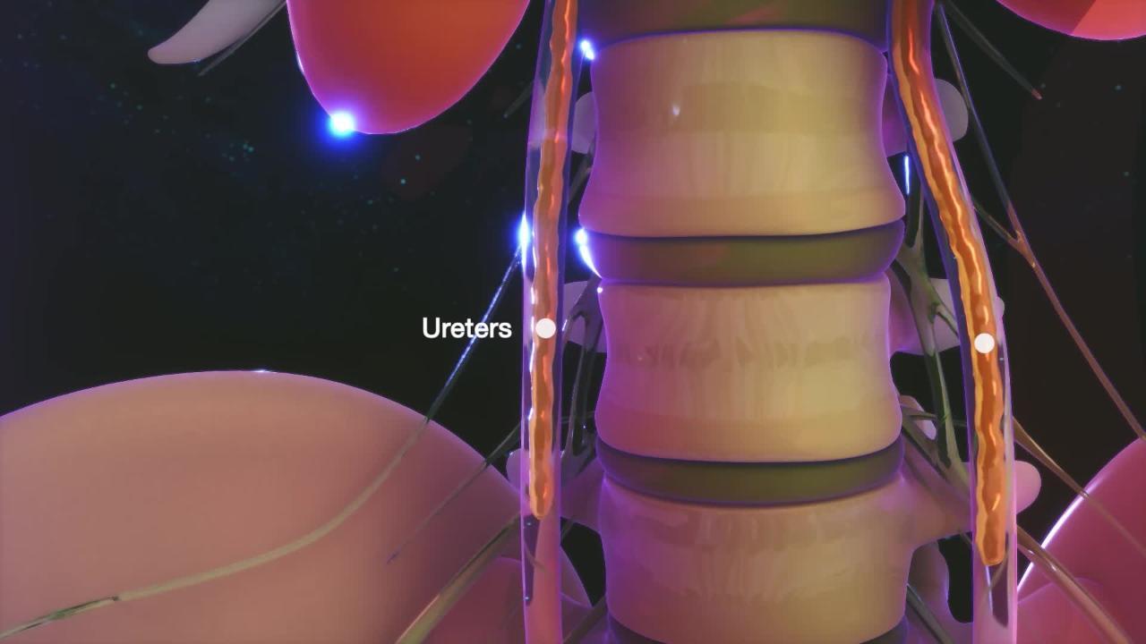
Cancer can occur in the cells lining the central collecting area of the kidney (the renal pelvis—usually the cancer is a type called urothelial carcinoma of the renal pelvis) and in the slender tubes that carry urine from the kidney to the bladder (ureters).
Cancers may cause blood in the urine or crampy pain in the side.
Diagnosis is usually by computed tomography.
Treatment is removal of the kidney and ureter.
Cancers of the renal pelvis and ureter are much less common than cancers of the rest of the kidney or bladder. They probably occur in fewer than 6,000 people in the United States each year.
Dalili za Saratani ya Figo ya Ureta na Fupanyonga
Blood in the urine is usually the first symptom. People may also have pain and burning during urination and an urgent, frequent need to urinate. Crampy pain in the flank (the space between the ribs and hip) or lower abdomen may occur if the flow of urine is obstructed (for example, because a blood clot blocks the ureter).
Utambuzi wa Saratani ya Figo ya Ureta na Fupanyonga
Computed tomography (CT) or ultrasound imaging
Ureteroscopy
The cancer is usually detected by using CT or ultrasound imaging. CT and often ultrasound can help doctors distinguish other noncancerous (benign) kidney and ureteral problems such as stones or blood clots. Microscopic examination of a urine sample may reveal cancer cells. A flexible viewing tube with a camera at its tip—a ureteroscope—threaded up through the bladder may be used to view cancers, obtain tissue samples for confirmation of the diagnosis, and occasionally even treat small cancers. This is typically done under general anesthesia. To determine how extensive cancers are and how far they have spread, CT scans of the abdomen and pelvis and chest x-ray or CT of the chest are done.
Matibabu ya Saratani ya Figo ya Ureta na Fupanyonga
Surgery
Sometimes chemotherapy before surgery
Sometimes laser therapy with medications
If the cancer has not spread beyond the area of the renal pelvis and ureter, the usual treatment is surgical removal of the entire kidney and ureter (nephroureterectomy) along with a small part of the bladder. Following surgery, chemotherapy is often instilled into the bladder to help prevent any recurrences in the bladder. However, in some situations—for example, when the kidneys are not functioning well or a person has only 1 kidney—the kidney is usually not removed because the person would then become dependent on dialysis.
For high-grade or high-stage tumors, chemotherapy is sometimes used before surgery.
Some cancers in the renal pelvis and ureter (for example, some low-grade and low-risk cancers) are treated with a laser to destroy the cancer cells or with surgery that removes only the cancer itself while leaving the kidney, the noncancerous portion of the ureter, and the bladder in place. However, these cancers have a higher risk of recurrence and spread. Occasionally, a medication, such as mitomycin C or bacille Calmette-Guérin (BCG—a substance that stimulates the body’s immune system), is instilled into the ureter or a chemotherapy medication is given. It is not clear how effective laser treatments and medication instillations are.
A cystoscopy (insertion of a flexible viewing tube to examine the inside of the bladder) is done periodically after surgery, indefinitely, because people who have had this type of cancer are at risk of developing bladder cancer.
Ubashiri wa Saratani ya Figo ya Ureta na Fupanyonga
If the cancer has not spread and if it can be completely removed surgically, cure is likely. However, if the cancer has spread into the wall of the renal pelvis or ureter or to distant sites, cure is unlikely.


