


The olecranon bursa lies immediately below the skin. This superficial location increases the risk of skin damage, fluid leakage, and infection from glucocorticoid injection. Thus, injection of glucocorticoids is usually avoided for superficial bursae. Occasionally, glucocorticoid injection is used to treat refractory or painful bursitis due to crystals (eg, gout) or rheumatoid arthritis, or if there is a significant recurrent noninfectious post-traumatic bursal effusion.
(See also Bursitis.)

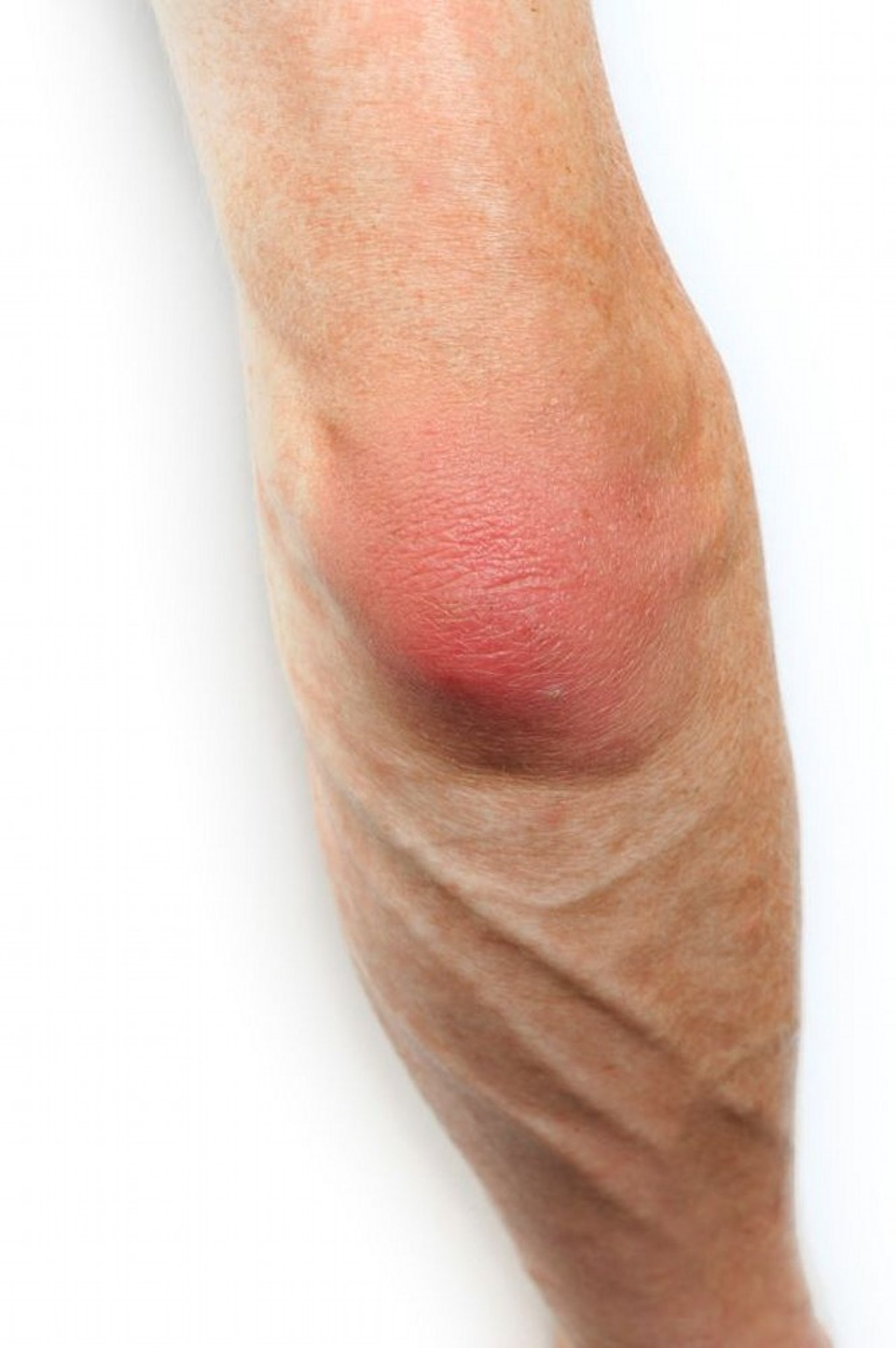
SCIENCE PHOTO LIBRARY
Indications for Aspirating or Injecting an Olecranon Bursa
Aspiration of bursal effusion to determine cause of bursitis
Rarely, injection of glucocorticoid for persistent or recurrent bursitis
Olecranon bursa aspiration is usually performed for diagnosis (eg, to diagnose septic or crystal-induced bursitis). The olecranon bursa is the most common site of septic bursitis; thus, an aspirated olecranon effusion should be sent to the laboratory for cell count and differential, crystal analysis, Gram stain, culture, and sensitivity tests (1).
Glucocorticoid injection is occasionally necessary in the olecranon bursa. Therapeutic injection should be performed only if all of the following criteria are satisfied:
Infection has been excluded by bursal fluid analysis.
Bursal fluid reaccumulates.
Symptoms are not relieved by local measures such as ice, elastic pressure bandage, and nonsteroidal anti-inflammatory drugs (NSAIDs) (if not contraindicated).
When needed, bursal injection may provide rapid relief, which can be particularly beneficial for large or painful effusions.
Contraindications to Aspirating or Injecting an Olecranon Bursa
Absolute contraindications
Hypersensitivity to an injected substance
For glucocorticoid injection, suspected septic bursitis
Insertion of a needle directly through infected skin should be avoided; however, if there is clinical suspicion for septic bursitis, the bursa should be aspirated, ideally before giving systemic antibiotics.
Relative contraindications
Poorly controlled diabetes: Any benefit of glucocorticoids is weighed against risk of worsening glycemic control and risk of infection.
Unresponsiveness to prior glucocorticoid injections into the same site (although this recommendation has not been systematically studied)
Coagulopathy is not a contraindication (2).
Complications of Aspirating or Injecting an Olecranon Bursa
Complications are uncommon and include:
Subcutaneous fat atrophy, skin atrophy and sinus tracts, temporary skin depigmentation, and infection.
Painful local reaction thought to result from a chemical bursitis in response to the crystals in the glucocorticoid solution (sometimes called postinjection flare) occurring within a few hours of depot glucocorticoid injection and usually lasting ≤ 48 hours
In diabetic patients, hyperglycemia after a depot glucocorticoid injection
Equipment for Aspirating or Injecting an Olecranon Bursa
Antiseptic solution (eg, chlorhexidine, povidone iodine, isopropyl alcohol)Antiseptic solution (eg, chlorhexidine, povidone iodine, isopropyl alcohol)
Sterile gauze and adhesive bandage
Gloves
20-mL syringe with 18- to 20-gauge, 1.5 inch needle for fluid withdrawal
Needle insertion site anesthesia: topical freezing spray (eg, ethyl chloride) (Needle insertion site anesthesia: topical freezing spray (eg, ethyl chloride) (3) or injectable 1% lidocaine without epinephrine, in a 3-mL syringe) or injectable 1% lidocaine without epinephrine, in a 3-mL syringe
Optional: For therapeutic injection, 5- to 10-mL syringe with 2 to 3 mL 1% lidocaine (without epinephrine) mixed with injectable depot glucocorticoid (eg, triamcinolone acetonide, 20 mg)Optional: For therapeutic injection, 5- to 10-mL syringe with 2 to 3 mL 1% lidocaine (without epinephrine) mixed with injectable depot glucocorticoid (eg, triamcinolone acetonide, 20 mg)
Hemostat, if switching of syringe while leaving the needle inserted after aspiration is anticipated
Some 3-, 5-, and 10-mL syringes
For diagnostic aspiration, appropriate tubes for specimen collection, including blood culture bottles
Having an assistant is helpful.
Additional Considerations for Aspirating or Injecting an Olecranon Bursa
For bursal injection, local anesthetic and depot glucocorticoid can be mixed in a single syringe. Adding the anesthetic helps confirm good needle placement when injection immediately relieves pain.
Glucocorticoid injection is rarely necessary in the olecranon bursa (based on an increased risk of infection and skin atrophy and a paucity of data showing improved long-term outcomes).
Septic bursitis cannot be ruled out by the initial gross and microscopic examination of the aspirated effusion; infected fluids (even from Staphylococcus aureus, the most common organism) tend to show a minimal fluid leukocytosis (although there is generally a high percentage of neutrophils). Hence, initial fluid analysis should be obtained prior to glucocorticoid injection. Septic bursitis requires drainage or rarely bursal excision in addition to systemic antibiotics.
Aspirated fluid is often serosanguinous.
Surgical excision of the bursa may be necessary for recalcitrant or recurrent sterile effusions or unresolving infections.
Relevant Anatomy for Aspirating or Injecting an Olecranon Bursa
The olecranon bursa overlies the tip of the olecranon process and is superficial.
Positioning for Aspirating or Injecting an Olecranon Bursa
Seat or partially recline the patient, with the arm comfortably flexed approximately 90° at the elbow and resting on a bedside table. The patient may also be supine on an examination table with the elbow flexed.
To avoid vasovagal episodes, avert the patient's head and orient your work area so that the patient does not see the needles.
Step-by-Step Description of Aspirating or Injecting an Olecranon Bursa
Prepare the site
Identify the bursa's point of maximum fullness and aspirate at the base (bottom) of the distended bursa, trying to avoid areas of skin thinning to limit likelihood of post-aspiration leakage.
Prepare the area with antiseptic solution.
Spray freezing solution at the needle insertion site until it just blanches or inject a skin wheal of local anesthetic (eg, ≤ 1 mL).
Puncture the bursa
Wear gloves (observe standard precautions).
Insert the needle (attached to the aspirating syringe) into the skin at the base of the bursa.
Advance the needle into the center of the bursa. Gently pull back on the plunger intermittently as you advance the needle tip to the center of the swelling.
Fluid will enter the syringe when the bursa is entered.
Drain all fluid from the bursa. Use your fingertips to apply gentle external pressure to the bursal sac to milk the fluid toward the needle tip.
If injecting the bursa, stabilize the needle hub with your hand and switch syringes. If the needle is on too tight, hold the hub of the needle with a hemostat.
Inject any medications and withdraw the needle.
Apply an adhesive bandage or sterile dressing.
Transfer bursal effusion samples to tubes and other transport media for fluid analysis. Inspect the fluid for blood and fat.
Aftercare for Aspirating or Injecting an Olecranon Bursa
A protective elastic elbow brace or compression bandage may prevent reaccumulation of fluid.
Prescribe limited activity; ice; and, if not contraindicated, oral nonsteroidal anti-inflammatory drugs (NSAIDs) until pain subsides.
Instruct the patient to return for reassessment to exclude infection if pain is continuously and progressively increasing after several hours or persists for > 48 hours, or if it resolves and then recurs.
Warnings and Common Errors for Aspirating or Injecting an Olecranon Bursa
Do not inject glucocorticoids against resistance; if there is resistance, slightly withdraw the needle.
Tips and Tricks for Aspirating or Injecting an Olecranon Bursa
Consider performing ultrasound if there is no obvious large effusion.
When inspecting bursal fluid, consider the following: The blood due to a traumatic needle insertion may be nonuniformly bloody and may clot. Fluid should be evaluated by polarized light microscopy for the presence of crystals.
References
1. Stell IM, Gransden WR. Simple tests for septic bursitis: comparative study. BMJ. 1998;316(7148):1877. doi:10.1136/bmj.316.7148.1877
2. Yui JC, Preskill C, Greenlund LS. Arthrocentesis and joint injection in patients receiving direct oral anticoagulants. Mayo Clin Proc 92(8):1223–1226, 2017. doi: 10.1016/j.mayocp.2017.04.007
3. Shah A, Vidoni A, McGarry S, et al. Ethyl chloride spray for musculoskeletal ultrasound-guided injections: An alternative to subcutaneous injection of local anesthetic solution. . Ethyl chloride spray for musculoskeletal ultrasound-guided injections: An alternative to subcutaneous injection of local anesthetic solution.J Clin Ultrasound. 2018;46(2):129-131. doi:10.1002/jcu.22561


