


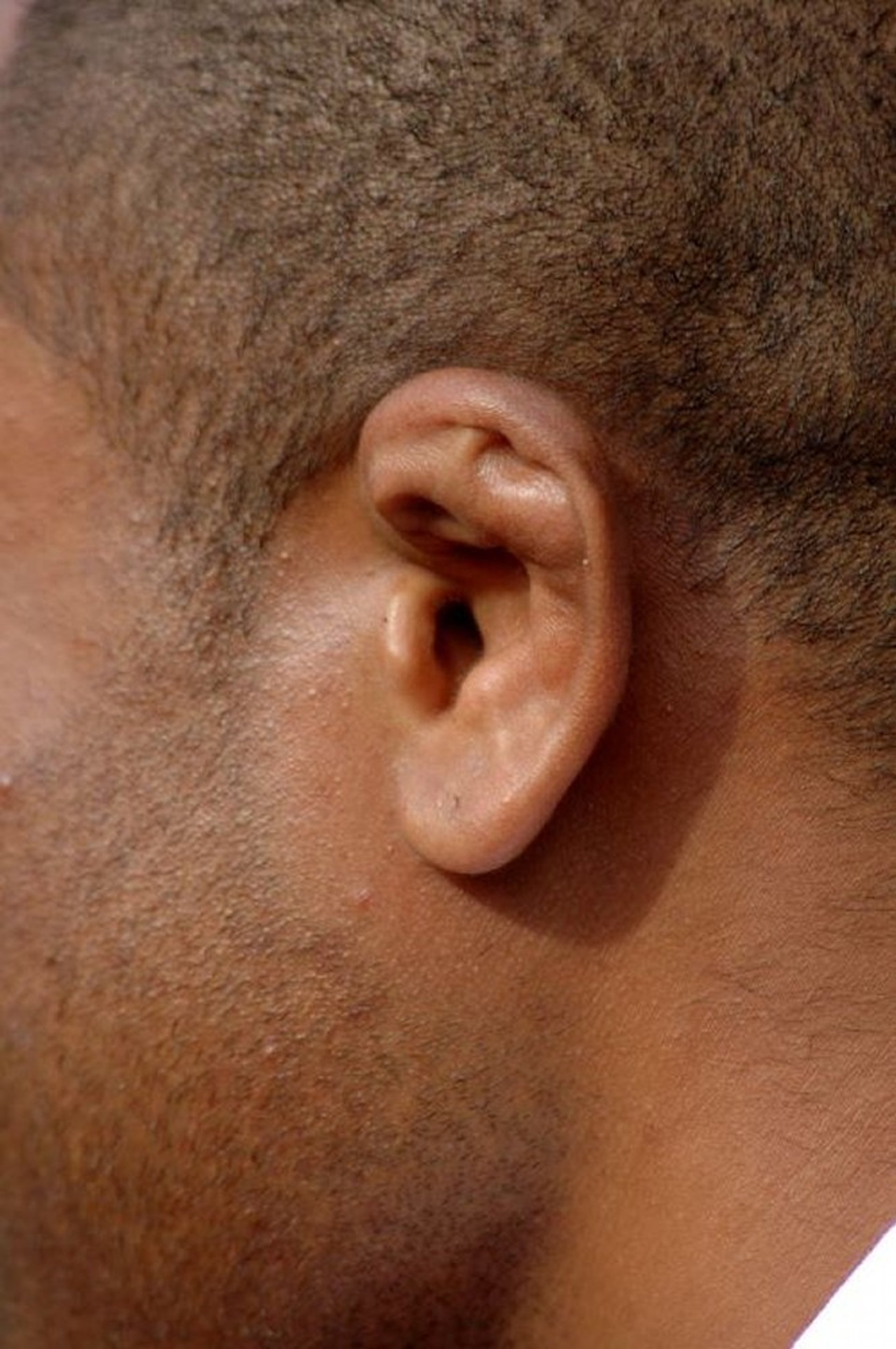
Auricular hematomas are drained to prevent chronic deformity of the underlying cartilage.
Auricular hematomas are caused by direct blunt trauma to the external ear, occurring most commonly in wrestlers, boxers, and rugby players. Trauma can cause a hematoma that separates the auricular perichondrium from the underlying cartilage and interrupts blood vessels that nourish the cartilage. If the perichondrium and its vessels are not reattached to the cartilage, there can be scarring and permanent deformity of the cartilage (cauliflower ear).
The goal of treatment is complete evacuation of the subperichondrial hematoma and replacement of the perichondrium back onto the cartilage surface to facilitate re-adhesion and prevent reaccumulation of the hematoma and cartilage malformation.
Needle aspiration of an auricular hematoma is no longer recommended due to the high risk of reaccumulation of the hematoma (1). This procedure focuses only on incision and drainage (2).

RICHARD WAREHAM FOTOGRAFIE/SCIENCE PHOTO LIBRARY
Indications for Draining an Auricular Hematoma
Tender focal swelling on the pinna within 7 days after trauma
If it has been longer than 7 days since the injury, the patient should be referred to an otolaryngologist for management of the ongoing perichondral inflammation and possible surgical intervention (evacuation or debridement) to prevent cartilaginous thickening.
Contraindications to Draining an Auricular Hematoma
Absolute contraindications
None
Relative contraindications
Cellulitis or other soft-tissue infections near the area of hematoma
Unrepaired laceration, particularly with exposed cartilage (repair the cartilage and laceration)
Chronic or recurrent hematoma (refer to an otolaryngologist)
Bleeding diatheses
Complications of Auricular Hematoma Drainage
Infection
Chondritis
Reaccumulation of the hematoma, which can lead to scar formation and if untreated, eventually cauliflower ear
Equipment for Auricular Hematoma Drainage
Sterile gloves
Antiseptic solution (eg, chlorhexidine, povidone iodine)Antiseptic solution (eg, chlorhexidine, povidone iodine)
Lidocaine 1%, without epinephrine, 3-mL syringe, 25-gauge needleLidocaine 1%, without epinephrine, 3-mL syringe, 25-gauge needle
Scalpel, #15
Hemostat or similar tool for dissection
Small suction
Syringe
Sterile normal saline
Optional: Sterile mineral oilOptional: Sterile mineral oil
Compression/bolster dressing material: Dental rolls, dry cotton, petrolatum gauze, or 4 × 4 plain gauze, gauze wrap dressing, and tape
Antibiotic ointment
Relevant Anatomy for Auricular Hematoma Drainage
Hematomas may be anterior, posterior, or both.
The skin of the auricle is normally densely adherent to the perichondrium, and if it is not held down with pressure, the cartilage below can necrose or deform over time.
The cartilage configuration of the auricle is relatively symmetric in most people, and this can help determine where to apply bolstering of a large anterior hematoma.


Positioning for Auricular Hematoma Drainage
For a posterior hematoma: Lateral decubitus position on the unaffected side
For an anterior hematoma: Supine with a slight head turn to the unaffected side
Step-by-Step Description for Draining an Auricular Hematoma
While wearing sterile gloves, cleanse the pinna and adjacent skin with antiseptic solution.
Anesthetize the pinna with 1% lidocaine without epinephrine using an Anesthetize the pinna with 1% lidocaine without epinephrine using anauricular block or direct infiltration around the hematoma. Topical anesthesia may be provided with lidocaine-prilocaine cream in addition or as an alternative method. or direct infiltration around the hematoma. Topical anesthesia may be provided with lidocaine-prilocaine cream in addition or as an alternative method.
Incise the skin along the posterior edge of the hematoma using the #15 scalpel blade. Follow the curvature of the pinna in a skin crease or at the edge of a change in the cartilage shape to hide the scar.
Gently separate the skin and overlying perichondrium from the hematoma and underlying cartilage using a hemostat or similar tool.
Manually evacuate the hematoma completely.
Irrigate the hematoma pocket with sterile normal saline.
Dry the area using gauze.
Apply antibiotic ointment to the incision edge.
Gently press the perichondrium back onto the cartilage with a finger; the pressure dressing will keep them in close proximity.
Place dry cotton into the external auricular canal.
Fill all external auricular crevices with petrolatum gauze, dental rolls, or gauze moistened with sterile saline or mineral oil.Fill all external auricular crevices with petrolatum gauze, dental rolls, or gauze moistened with sterile saline or mineral oil.
Hematomas that are both anterior and posterior require the bolsters to be sutured together with monofilament suture; dental rolls are particularly suited for this maneuver.
Place several layers of gauze behind the ear to support the back of the ear against the pressure dressing. Cut a V-shape or curve out of the gauze first to allow for a close fit to the ear.
Cover the external ear with multiple layers of fluffed-up 4 × 4 gauze, and hold them in place by wrapping elastic or gauze bandages around the head and taping the bottom edge of the dressing over the ear lobule.
Aftercare for Auricular Hematoma Drainage
Prescribe oral antibiotics (eg, cephalexin) effective against staphylococcus for 7 days.Prescribe oral antibiotics (eg, cephalexin) effective against staphylococcus for 7 days.
The dressing should be kept dry for 1 week; a shower cap should be used when bathing.
Reevaluate for recurrence of the hematoma 24 hours after removal of the dressing.
Remove the dressing after 1 week.
Patients should not manipulate the ear for 1 month after the dressing is removed to optimize healing and cosmetic results.
Warnings and Common Errors for Auricular Hematoma Drainage
The pressure dressing must be snug enough to prevent hematoma recurrence and/or fluid accumulation, but not so tight as to impede circulation. Typically, if the patient feels the dressing is increasing the pain or causing a headache, the dressing is too tight and should be loosened. If the patient does not feel pressure on the ear after the wrap is placed, it is too loose.
Tips and Tricks for Auricular Hematoma Drainage
Ensure adequate anesthesia is administered for proper positioning of the perichondrium after hematoma evacuation.
The cotton or gauze bolsters should be placed in small pieces to conform precisely to the shape of the ear cartilage. This presses the perichondrium firmly against the cartilage, allowing it to heal without reaccumulation of the hematoma or deformity.
A properly applied head wrapping is necessary to prevent its impinging uncomfortably on the patient's eyebrow and contralateral ear. This can be mitigated by wrapping umbilical tape or an unfolded 4 × 4 gauze around the dressing above the ipsilateral eye and the opposite ear to prevent the head wrapping from pressing on the eyebrow or opposite ear.
References
1. Giles WC, Iverson KC, King JD, et al. Incision and drainage followed by mattress suture repair of auricular hematoma. Laryngoscope. 2007;117(12):2097-2099. doi:10.1097/MLG.0b013e318145386c
2. Long B, Mason J, Bridwell RE, et al. Managing Auricular Hematoma: An Emergency Medicine Narrative Review. J Emerg Med. 2025;69:62-75. doi:10.1016/j.jemermed.2024.08.021


