


An auricular block anesthetizes the pinna.
Topic Resources

Indications for Auricular Block
Because there is minimal subcutaneous tissue on the pinna, direct injection of local anesthetic to anesthetize the pinna can be difficult and uncomfortable. Auricular blocks may be used for pain relief prior to:
Repair of laceration of the pinna or excision of minor auricular lesions
Drainage of an auricular hematoma
Auricular neuralgia (eg, due to herpetic lesions)
Contraindications to Auricular Block
Absolute contraindications:
None
Relative contraindications:
Infection in the path of needle insertion: Use procedural sedation or other anesthesia
History of known hypersensitivity to the anesthetic agent or delivery vehicle (choose a different anesthetic)
Complications of Auricular Block
Adverse reaction to the anesthetic or delivery vehicle (eg, known hypersensitivity reaction to the anesthetic [rare] or to methylparaben [a preservative])
Rarely, systemic toxicity due to anesthetic overdose (eg, seizure, cardiac arrhythmias), or inadvertent systemic absorption of anesthetic (if injected into a blood vessel)
Hematoma
Horner syndrome, if the local anesthetic spreads to the sympathetic chain, leading to ptosis, miosis, and anhidrosis; it is usually transient and resolves spontaneously
Equipment for Auricular Block
Nonsterile gloves, mask, and safety glasses or a face shield
Antiseptic solution (eg, chlorhexidine, povidone iodine)Antiseptic solution (eg, chlorhexidine, povidone iodine)
Injectable local anesthetic such as lidocaine 2% with or without epinephrine* 1:100,000 or, for longer-duration anesthesia, bupivacaine 0.5% with or without epinephrine* 1:200,000Injectable local anesthetic such as lidocaine 2% with or without epinephrine* 1:100,000 or, for longer-duration anesthesia, bupivacaine 0.5% with or without epinephrine* 1:200,000
5- to 10-mL syringe, 25- or 27-gauge needle: 3-cm long for injection
*Maximum dose of local anesthetics: Lidocaine without epinephrine, 5 mg/kg; lidocaine with epinephrine, 7 mg/kg; bupivacaine, 1.5 mg/kg. NOTE: A 1% solution (of any substance) represents 10 mg/mL (1 gm/100 mL). Epinephrine causes vasoconstriction, which prolongs the anesthetic effect. Patients with cardiac disease should receive only limited amounts of epinephrine (maximum 3.5 mL of solution containing 1:100,000 epinephrine); alternatively, use local anesthetic without epinephrine.*Maximum dose of local anesthetics: Lidocaine without epinephrine, 5 mg/kg; lidocaine with epinephrine, 7 mg/kg; bupivacaine, 1.5 mg/kg. NOTE: A 1% solution (of any substance) represents 10 mg/mL (1 gm/100 mL). Epinephrine causes vasoconstriction, which prolongs the anesthetic effect. Patients with cardiac disease should receive only limited amounts of epinephrine (maximum 3.5 mL of solution containing 1:100,000 epinephrine); alternatively, use local anesthetic without epinephrine.
Additional Considerations for Auricular Block
Consider using sedation or an alternative method of anesthesia for patients unable to cooperate with the procedure.
Relevant Anatomy for Auricular Block
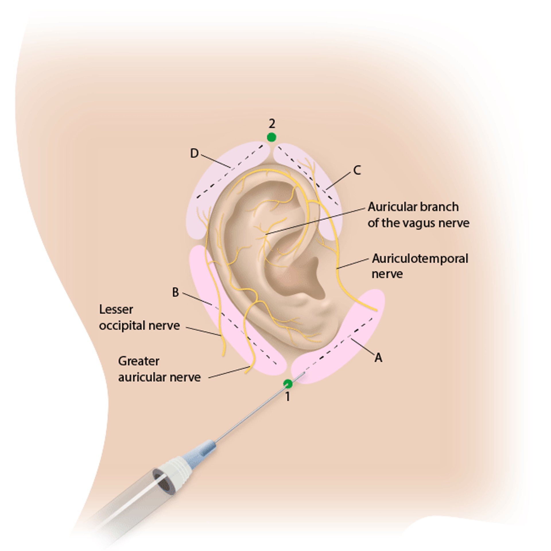
The auricle is innervated by 3 nerves:
Greater auricular nerve
Lesser occipital nerve
Auriculotemporal nerve
The relevant anatomic landmarks of the ear are the helical root, concha, lobule.
Positioning for Auricular Block
Supine with the head supported on a pillow and rotated so the affected ear is upward
Step-by-Step Description of Auricular Block
See also figure Auricular Block.
Apply nonsterile gloves, mask, and safety glasses or a face shield.
Cleanse ear and surrounding skin with antiseptic solution and allow to dry.
There are 2 needle insertion sites: one approximately 2 cm inferior to the lobe of the ear (see 1 on figure Auricular Block) and the other approximately 1 cm superior to the pinna (see 2 on figure Auricular Block).
Inject a small amount of anesthetic at the injection site inferior to the lobe (see 1 on figure Auricular Block) to form a skin wheal. Direct the needle toward a point 1 cm anterior to the tragus (see A on figure Auricular Block) and inject 2 mL of anesthetic as the needle is withdrawn. Do not completely remove the needle from the subcutaneous space.
Redirect the needle toward the mastoid process (see B on figure Auricular Block) and inject 2 mL of anesthetic as the needle is withdrawn.
Inject a small amount of anesthetic superior to the pinna (see 2 on figure Auricular Block) to form a skin wheal. Direct the needle toward a point 1 cm anterior to the tragus (see C on figure Auricular Block) and inject 2 mL of anesthetic as the needle is withdrawn. Do not completely remove the needle from the subcutaneous space.
Redirect the needle toward the mastoid process (see D on figure Auricular Block) and inject 2 mL of anesthetic as the needle is withdrawn.
Await onset of anesthesia (3 to 5 minutes).
Auricular Block

Aftercare for Auricular Block
Ensure hemostasis at the injection site.
Instruct patient regarding anticipated time to anesthesia resolution.
Additional care based on the procedure performed after nerve block was completed.
Tips and Tricks for Auricular Block
Distraction techniques (eg, talking to the patient or having the patient hold someone else's hand) may help to reduce patient anxiety.
Inject the local anesthetic solution slowly (30 to 60 seconds) to reduce the pain of injection.


