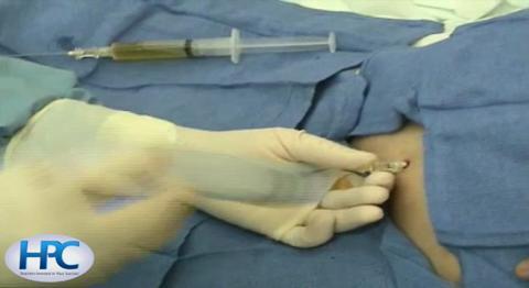
Paracentesis is removal of peritoneal fluid (ascites or ascitic fluid) from the abdomen with percutaneous needle aspiration.
Ресурси за темою
Paracentesis can be done for diagnosis, to analyze ascitic fluid (in which small quantities are removed), or for treatment, typically in patients with chronic tense ascites (in which case large quantities are removed).
(See also Paracentesis.)
Indications for Paracentesis
Diagnostic paracentesis
In patients who have peritoneal fluid that is new or of uncertain etiology
In patients with ascites and symptoms such as fever or increased pain that suggest possible infection of the ascitic fluid (eg, spontaneous bacterial peritonitis)
Therapeutic paracentesis
To alleviate symptoms, usually dyspnea or pain caused by large-volume ascites
Many causes of peritonitis are surgical emergencies and do not require paracentesis.
Selection of laboratory tests typically done on ascitic fluid is discussed in diagnosis of ascites.
Contraindications to Paracentesis
Absolute contraindications
Severe, uncorrectable disorders of blood coagulation
Intestinal obstruction with bowel distention (unless an area of peritoneal fluid that can be safely entered has been identified using imaging studies)
An infected abdominal wall
Relative contraindications
Poor patient cooperation
Surgical scarring at the puncture site: The paracentesis site should be moved away from any area of scarring. Surgical scars can cause adherence of the bowel to the abdominal wall, increasing the risk of bowel perforation during paracentesis.
Large intra-abdominal mass, or second or third trimester pregnancy: In these patients, paracentesis should be done with ultrasound guidance.
Severe portal hypertension with abdominal collateral circulation: This disorder increases the risk of needle injury to dilated veins in the abdominal wall.
Complications of Paracentesis
Hemorrhage due to needle injury to an artery or vein: Intra-abdominal bleeding can be difficult to control and can be fatal.
Prolonged leakage of ascitic fluid through the needle puncture site
Infection (eg, due to contamination by the needle or skin flora)
Bowel perforation, resulting in leakage of bowel contents into the peritoneum and infection of the ascitic fluid
With large-volume paracentesis, hypotension and possibly transient hyponatremia and increased creatinine
Equipment for Paracentesis
Signed consent form
Local anesthetic (eg, 10 mL of 1% lidocaine), 25- and 20- to 22-gauge needles, and 10-mL syringe
Antiseptic solution with applicators, drapes, and gloves
Sterile gauze sponges
Paracentesis needle, such as an 18- to 22-gauge (1.5-inch or 3.5-inch as needed) needle for diagnostic paracentesis, an 18- to 14-gauge (1.5-inch or 3.5-inch as needed) or a 15-gauge (3.25-inch) Caldwell needle with overlying metal catheter for therapeutic paracentesis
#11 scalpel blade (may be needed to widen the entry site, particularly for large-volume paracentesis and larger needles)
3-way stopcock
30- to 50-mL syringe
Wound dressing materials (eg, adhesive bandages)
Appropriate containers (eg, red top and purple top tubes, blood culture bottles) for collection of fluid for laboratory tests
For removal of large volumes, vacuum bottles or collection bags
If ultrasound guidance is used, ultrasonographic equipment
Additional Considerations for Paracentesis
Colloid replacement, such as concurrent infusion of IV albumin (6 to 8 g/L of ascitic fluid removed or 50 g) or dextran-70 (which has no infection risk), is sometimes recommended during large-volume paracentesis (eg, removal of > 5 L) to help avoid significant intravascular volume shift and post-procedure hypotension.
If ultrasound-guided paracentesis is done, once the site is marked, the procedure should be done with real-time guidance or the patient should be kept immobile and the paracentesis done as soon as possible to avoid shifting of the fluid or intra-abdominal organs.
Ultrasonographic guidance should be used whenever preferred by the operator, during the second or third trimester of pregnancy, with a large intra-abdominal mass, or when there is a scar. If there is a scar, the paracentesis can be done blindly at a location away from the scar.
Positioning for Paracentesis
Have the patient sit in bed with the head elevated 45 to 90°. If choosing a needle insertion site in the left lower quadrant, partially roll the patient onto his or her left side to allow the fluid to pool in the area.
Alternatively, position the patient in a lateral decubitus position. In this position, the air-filled bowel loops float up.
Relevant Anatomy for Paracentesis
The linea alba is the midline fibrous band the runs vertically from the xiphoid process to the pubic symphysis. This fibrous band does not contain important nerves or blood vessels.
Step-by-Step Description of Paracentesis
Explain the procedure to the patient and obtain written informed consent.
Ask the patient to empty the bladder by voiding, or catheterize the patient.
Place the patient in bed with the head elevated 45 to 90°. In patients with obvious and a large amount of ascites, locate an insertion site at the midline between the umbilicus and the pubic bone, about 2 cm below the umbilicus. Locate an alternative site in the left lower quadrant, eg, about 3 to 5 cm superior and medial to the anterior superior iliac spine. If choosing the left lower quadrant site, roll the patient partially onto the left side to allow the fluid to pool in the area. The insertion site should be lateral enough to avoid the rectus sheath, which contains the inferior epigastric artery.
Alternatively, place the patient in a lateral decubitus position. In this position, the air-filled bowel loops float up, migrating away from the point of entry, which should be down in the fluid-filled region. The left lateral decubitus position with needle insertion in the left lower quadrant is preferred by some physicians because the cecum may be distended with gas in the right lower quadrant. The right lateral decubitus position can be used if needed.
To choose a needle insertion site, carefully percuss, because dullness to percussion confirms the presence of fluid.
If needed, use ultrasound to identify a site, confirming the presence of ascitic fluid and the absence of overlying bowel.
In selecting an insertion site, avoid surgical scars and visible veins.
If available, mark the insertion site with a skin marking pen.
Prepare the area with a skin cleansing agent, such as chlorhexidine or povidone iodine, and apply a sterile drape while wearing sterile gloves.
Using a 25-gauge needle, place a wheal of local anesthetic over the insertion point. Switch to a larger (20- or 22-gauge) needle and inject anesthetic progressively deeper until reaching the peritoneum, which should also be infiltrated because it is sensitive. When the needle is advanced, maintain constant negative pressure to ensure lidocaine is not injected into a blood vessel.
For diagnostic paracentesis, select an 18- to 22-gauge (1.5-inch or 3.5-inch as needed) needle. For therapeutic paracentesis, select an 18- to 14-gauge (1.5-inch or 3.5-inch as needed) needle or a Caldwell needle (15-gauge, 3.25-inch). Smaller-gauge needles lessen the risk of complications, such as ascitic fluid leakage, but take longer to complete therapeutic paracentesis.
Insert the needle perpendicular to the skin at the marked site. Alternatively, insert the needle using the Z-track method, which can be done in several ways. One option: Pull the skin, insert the needle perpendicularly, and maintain this skin traction until the needle enters the peritoneal cavity. Another option: Puncture just the skin, and pull it down, then advance into the peritoneal cavity. A third option: Insert the needle at an angle (usually 45°) to the skin and advance it. The Z-track method is preferred because it allows the intra-abdominal pressure to help seal the tract after removing the needle and decreases the risk of peritoneal fluid leak.
Insert the needle slowly to help avoid puncturing the bowel and use intermittent suction to avoid entering into a blood vessel. Avoid continuous suction because this can cause tissue (eg, bowel, omentum) to occlude the needle tip.
Insert the needle through the peritoneum (generally accompanied by a popping sensation) and gently aspirate fluid.
For diagnostic paracentesis, withdraw enough fluid (eg, 30 to 50 mL) into the syringe and place the fluid in appropriate tubes and bottles for testing, including blood culture bottles.
For therapeutic paracentesis, if a Caldwell needle is used, advance the outer metal catheter over the needle, then remove the needle from inside the catheter. Attach the catheter to a collection bag or vacuum bottle using tubing.
For therapeutic paracentesis, a large volume of fluid is removed. Removal of 5 to 6 L of fluid is generally well tolerated. In some patients, up to 8 L can be removed. Colloid replacement, such as concurrent infusion of IV albumin, is often recommended during large-volume paracentesis (eg, removal of > 5 L) to help avoid significant intravascular volume shift and post-procedure hypotension.
A 3-way stopcock can be used to control the flow of fluid when changing collection bottles or if a diagnostic sample is needed.
Remove the needle and apply pressure to the site.
Apply a sterile adhesive bandage to the insertion site.
Aftercare for Paracentesis
If there is significant leakage of ascitic fluid, apply a pressure bandage.
After large volume paracentesis, monitor blood pressure for 2 to 4 hours after the procedure.
Warnings and Common Errors for Paracentesis
Before needle insertion, there must be dullness to percussion to confirm the presence of fluid and lack of overlying bowel. If not certain, use ultrasound to identify a site, confirming the presence of ascitic fluid and the lack of overlying bowel.
Tips and Tricks of Paracentesis
If flow of ascitic fluid stops during paracentesis, gently rotate the needle or catheter and advance in 1- to 2-mm increments. If the flow does not resume, briefly release suction from the vacuum (usually using the 3-way stopcock) and then resume suctioning. You can alternatively slowly withdraw the catheter in 1- to 2-mm increments, but once out of the peritoneum, the catheter cannot be reinserted, so this should be done cautiously.
Some patients require repeated paracentesis. Use this previous experience as a guide to locate the insertion site and estimate the amount of fluid that can safely be removed.

