


Atelectasis refers to the collapse of lung tissue with loss of volume. Patients may have dyspnea or respiratory failure if atelectasis is extensive. They may also develop pneumonia. Atelectasis is usually asymptomatic, but hypoxemia and pleuritic chest pain may be present. Diagnosis is by chest radiography or computed tomography. Treatment includes coughing, deep breathing, and treating the cause.
Topic Resources
")
The natural tendency for open air spaces such as the alveoli to collapse is countered by the following:
Surfactant (which maintains low surface tension)
Continuous breathing (which keeps the alveoli open)
Intermittent deep breathing (which releases surfactant into the alveoli)
Periodic coughing (which clears the airways of secretions)
Major consequences of atelectasis include impaired gas exchange (ie, hypoxia and/or hypercapnia), pneumonia, and respiratory failure in severe cases.
Etiology of Atelectasis
The most common factors that can cause atelectasis include the following:
Intrinsic obstruction of airways (eg, by foreign body, tumor, mucous plug)
Extrinsic compression of airways (eg, by tumors, lymphadenopathy)
Suppression of respiration or cough (eg, by general anesthesia, oversedation, pain)
Supine positioning, particularly in patients with obesity or cardiomegaly
Compression or collapse of lung parenchyma (eg by large pleural effusion or pneumothorax)
Thoracic and abdominal surgeries are very common causes because they involve general anesthesia, opioid use (with possible secondary respiratory depression), and often painful respiration. A malpositioned endotracheal tube can cause atelectasis by occluding a mainstem bronchus.
Less common causes of atelectasis include surfactant deficiency or dysfunction, lung parenchymal scarring, or tumors.
Symptoms and Signs of Atelectasis
Atelectasis itself is asymptomatic unless hypoxemia or pneumonia develops. Symptoms of hypoxemia tend to be related to the acuity of onset and extent of atelectasis. With rapid, extensive atelectasis, dyspnea or even respiratory failure can develop. With slowly developing, less extensive atelectasis, symptoms may be mild or absent.
Pneumonia may cause cough, dyspnea, and pleuritic pain. Pleuritic pain may also be due to the disorder that initially caused atelectasis (eg, chest trauma, surgery).
Signs are often absent if atelectasis is minimal. Decreased breath sounds in the region of atelectasis and possibly dullness to percussion and decreased chest excursion are detectable if the area of atelectasis is large. Often it is discovered incidentally on chest imaging (eg, radiographs or computed tomography [CT]).
Diagnosis of Atelectasis
Chest radiograph or, particularly in more mild disease, chest CT
Atelectasis should be suspected in patients who have any unexplained respiratory symptoms and who have risk factors, particularly recent major surgery. Atelectasis that is clinically significant (eg, that causes symptoms, increases risk of complications, or meaningfully affects pulmonary function) is generally visible on chest radiographs; findings can include localized lung opacification and/or loss of lung volume. More mild cases may not be apparent on chest radiographs and can only be seen with cross-sectional imaging (ie chest CT).

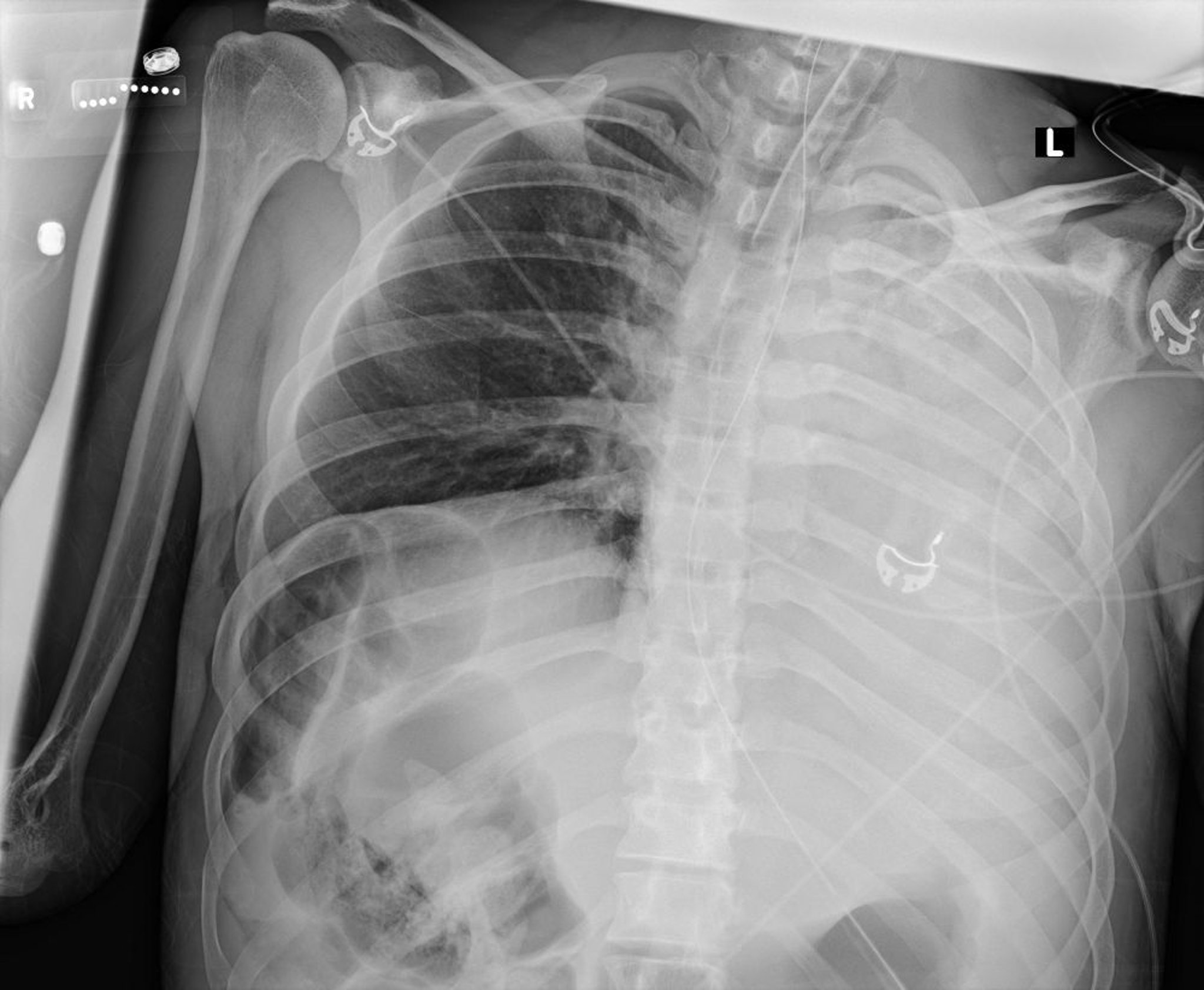
Courtesy of Alexander S. Niven, MD
If the cause of atelectasis is not clinically apparent (eg, if it is not recent surgery or pneumonia is not identified on chest radiographs) or another disorder is suspected (eg, pulmonary embolism, tumor), other tests, such as bronchoscopy or chest computed tomography (CT), may be necessary.
Treatment of Atelectasis
Maximizing cough and deep breathing
If obstruction by tumor or foreign body is suspected, bronchoscopy
Evidence for the efficacy of most treatments for atelectasis is weak. Nonetheless, commonly recommended measures include chest physiotherapy to help maintain ventilation and clearance of secretions, and encouragement of lung expansion techniques such as directed cough, deep breathing exercises, and frequent use of an incentive spirometer, which is usually done in inpatient settings (1). In ambulatory patients, exercise (eg, walking) is a desirable way to promote deep breathing.
For patients who are intubated and mechanically ventilated, positive end-expiratory pressure may prevent atelectasis. While higher tidal volumes may reduce atelectasis, evidence supports use of lower volumes (eg, 6 to 8 cm H2O) for the vast majority of patients to reduce the risk of ventilator-induced lung injury. For patients who are not intubated and do not have excessive secretions, continuous positive airway pressure may help (2).
Avoiding oversedation helps ensure ventilation and sufficient deep breathing and coughing. However, severe pleuritic pain may impair deep breathing, and coughing and may be relieved only with opioids. Thus, many clinicians prescribe opioid analgesics in doses sufficient to relieve pain and advise patients to consciously cough and take deep breaths periodically. In certain postoperative patients, epidural analgesia or an intercostal nerve block may be used to relieve pain without causing respiratory depression. Antitussive therapy should be avoided.
Most importantly, the underlying cause of atelectasis (eg, mucous plug, foreign body, tumor, mass, pulmonary effusion) should be treated. For persistent mucous plugging, nebulized dornase alfa (which breaks down DNA in mucus) and sometimes bronchodilators are tried; both help with liquefaction and expectoration of tenacious mucus (Most importantly, the underlying cause of atelectasis (eg, mucous plug, foreign body, tumor, mass, pulmonary effusion) should be treated. For persistent mucous plugging, nebulized dornase alfa (which breaks down DNA in mucus) and sometimes bronchodilators are tried; both help with liquefaction and expectoration of tenacious mucus (3). Acetylcysteine is effective but sometimes avoided because it can cause bronchoconstriction. If other measures are ineffective or if a cause of obstruction other than mucous plugging is suspected, bronchoscopy should be performed.). Acetylcysteine is effective but sometimes avoided because it can cause bronchoconstriction. If other measures are ineffective or if a cause of obstruction other than mucous plugging is suspected, bronchoscopy should be performed.
Treatment references
1. Odor PM, Bampoe S, Gilhooly D, Creagh-Brown B, Moonesinghe SR. Perioperative interventions for prevention of postoperative pulmonary complications: systematic review and meta-analysis. BMJ 2020;368:m540. Published 2020 Mar 11. doi:10.1136/bmj.m540
2. Branson RD. The scientific basis for postoperative respiratory care. Respir Care 2013;58(11):1974-1984. doi:10.4187/respcare.02832
3. Tarrant BJ, Maitre CL, Romero L, et al. Mucoactive agents for adults with acute lung conditions: A systematic review. Heart Lung 2019;48(2):141-147. doi:10.1016/j.hrtlng.2018.09.010
Prevention of Atelectasis
People who smoke can decrease their risk of postoperative atelectasis by stopping smoking, ideally at least 6 to 8 weeks before surgery. Pharmacotherapy for patients with chronic lung disorders (eg, chronic obstructive pulmonary disease [COPD]) should be optimized before surgery.
Preoperative inspiratory muscle training (including incentive spirometry) should be considered for patients scheduled for thoracic or upper abdominal surgery. During surgery or in the immediate perioperative period, using prophylactic respiratory physiotherapy, lung-protective ventilation modes and postoperative continuous positive airway pressure (by providing constant expiratory pressure and preventing alveolar collapse) have all been helpful in preventing atelectasis. After surgery, early ambulation and lung expansion techniques (eg, coughing, deep breathing exercises, incentive spirometry) may also decrease risk.
Key Points
Atelectasis is reversible collapse of lung tissue with loss of volume; common causes include intrinsic or extrinsic airway compression, hypoventilation, and a malpositioned endotracheal tube.
A large area of atelectasis may cause symptomatic hypoxemia, but any other symptoms are due to the cause or a superimposed pneumonia.
Diagnosis is by chest radiography; if the cause is not clinically apparent, bronchoscopy or chest computed tomography may be needed.
Treatment involves using incentive spirometry, maximizing coughing, deep breathing, and, whenever possible, encouraging mobilization.


