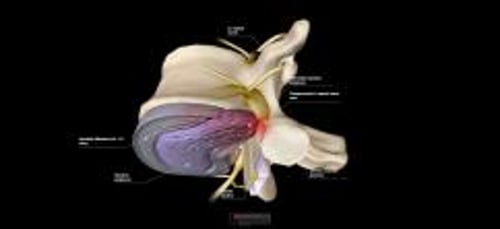
Herniated lumbar disc is prolapse of the nucleus pulposus of an intervertebral disc through a tear in the surrounding annulus fibrosus. The tear causes pain due to irritation of sensory nerves in the disc, and when the disc impinges on an adjacent nerve root, a segmental radiculopathy with paresthesias and sometimes weakness in the distribution of the affected root results. Diagnosis is usually confirmed by MRI or CT. Treatment of mild cases is with analgesics, activity modification, and physical therapy. Patients with progressive or severe neurologic deficits, intractable pain, conservative treatment failure, myelopathy, or cauda equina syndrome with associated sphincter dysfunction may require immediate or later elective surgery (eg, diskectomy, laminectomy).
Spinal vertebrae are separated by fibrocartilaginous discs consisting of an outer annulus fibrosus and an inner nucleus pulposus. When degenerative changes (with or without trauma) result in protrusion or rupture of the nucleus through the annulus fibrosus in the lumbosacral or cervical area, the nucleus is displaced posterolaterally or posteriorly into the extradural space.
Radiculopathy occurs when the herniated nucleus compresses or irritates the nerve root. Posterior protrusion may compress the cord in the cervical, thoracic, or upper lumbar spine or the cauda equina, especially in a congenitally narrow spinal canal (spinal stenosis). In the lumbar area, the majority of disc ruptures affect L5 or S1 nerve roots.
Herniated discs are common.
Symptoms and Signs of Lumbar Disc Herniation
Herniated discs often cause no symptoms, or they may cause symptoms and signs in the distribution of affected nerve roots. Pain usually develops suddenly, and back pain is typically relieved by rest and activity modification. In contrast, nerve root pain caused by an epidural tumor or abscess begins more insidiously, and back pain is often worse at night while in bed.
Symptomatic cauda equina compression often results in urine retention or incontinence due to loss of sphincter function.
In patients with lumbosacral disc herniation, coughing, sneezing, and activities such as sitting and bending forward increase the pain.
© Springer Science+Business Media
Diagnosis of Lumbar Disc Herniation
MRI or CT
Diagnosis is usually suspected during a history and physical examination and confirmed by MRI or CT.
The physical examination should include the an evaluation of strength, sensation, and reflexes. Tests for dural tension should also be done.
On examination, with the patient in supine position with legs extended, raising the leg may cause pain to radiate down the posterior thigh to below the knee (straight leg-raising test). Pain can be bilateral with central disc herniation.
Straightening the knee while sitting can produce similar pain (sitting straight-leg raising test). A variation of this test in which straightening the knee with the patient sitting and bending forward at the waist with the foot dorsiflexed is called the slump test.
In upper lumbar disc herniation (L1-2, L2-3), extending the leg at the hip with the patient prone can cause pain radiating into the anterior thigh (femoral stretch test).
Achilles tendon and patellar reflexes may be diminished or absent.
MRI or CT can identify the cause and precise level of the lesion. Rarely (ie, when MRI is contraindicated and CT is inconclusive), CT myelography is necessary. Electrodiagnostic testing may help identify the involved root if the pain has been present for at least 2 weeks. Earlier testing may result in a false-negative exam.
Because an asymptomatic herniated disc is common, the clinician must carefully correlate symptoms and physical examination findings with MRI abnormalities before invasive procedures are considered.
Treatment of Lumbar Disc Herniation
Conservative treatment initially
Invasive procedures, sometimes including surgery, if neurologic deficits are progressive or severe
Because a herniated disc desiccates and will reabsorb within 6 to 9 months (1), symptoms tend to abate regardless of treatment. Up to 85% of patients with back pain—regardless of cause—recover without surgery within 6 weeks (2).
Conservative treatment
Treatment of a herniated disc should be conservative unless neurologic deficits are progressive or severe (3). Heavy or vigorous physical activity is restricted, but ambulation and light activity (eg, lifting objects < 2.5 to 5 kg [about 5 to 10 lb] using correct techniques) are permitted as tolerated; prolonged bed rest and traction are not indicated.
Physical therapy and home exercises can improve posture and strengthen back muscles and thus reduce spinal movements that further irritate or compress the nerve root.
Surgical procedures
Surgical procedures should be considered for the following:
Persistent or worsening neurologic deficits, particularly objective deficits (eg, weakness, reflex deficits) due to cervical or lumbar radiculopathies
Acute compression of the spinal cord or cauda equina syndrome
Severe, intractable nerve root pain or sensory deficits
Immediate surgical evaluation is needed if clinical findings of spinal cord compression correlate with MRI abnormalities.
Microscopic diskectomy and laminectomy with surgical removal of herniated material are usually the procedures of choice. Percutaneous approaches to remove bulging disc material are still being evaluated.
Dissolving herniated disc material with local injections of the enzyme chymopapain is not recommended.
Lesions acutely compressing the spinal cord or cauda equina (eg, causing urine retention or incontinence) require immediate surgical evaluation (see diagnosis of spinal cord compression).
Treatment references
1. Zou T, Liu XY, Wang PC, et al. Incidence of Spontaneous Resorption of Lumbar Disc Herniation: A Meta-analysis. Clin Spine Surg. 2024;37(6):256-269. doi:10.1097/BSD.0000000000001490
2. Kreiner DS, Hwang SW, Easa JE, et al. An evidence-based clinical guideline for the diagnosis and treatment of lumbar disc herniation with radiculopathy. Spine J. 2014;14(1):180-191. doi:10.1016/j.spinee.2013.08.003
3. Qaseem A, Wilt TJ, McLean RM, et al. Noninvasive Treatments for Acute, Subacute, and Chronic Low Back Pain: A Clinical Practice Guideline From the American College of Physicians. Ann Intern Med. 2017;166(7):514-530. doi:10.7326/M16-2367
4. Chou R, Pinto RZ, Fu R, et al. Systemic corticosteroids for radicular and non-radicular low back pain. Cochrane Database Syst Rev. 2022;10(10):CD012450. Published 2022 Oct 21. doi:10.1002/14651858.CD012450.pub2
Key Points
Herniated lumbar discs are common and usually affect nerve roots at L5 or S1.
Because most disc herniations reabsorb within 6 to 9 months, symptoms tend to improve regardless of treatment.
Diagnose based on history and physical examination findings, and confirm by MRI or CT.
If symptoms develop suddenly and back pain is relieved with rest, suspect a herniated disc.
Recommend analgesics, light activity as tolerated, and exercises to improve posture and strength; however, if pain or deficits are severe or worsening, consider surgical procedures.

