

Benign liver tumors are relatively common. Most are asymptomatic, but some cause hepatomegaly, right upper quadrant discomfort, or intraperitoneal hemorrhage. Most are detected incidentally on ultrasound or other scans (see Imaging Tests of the Liver and Gallbladder). Liver tests are usually normal or only slightly abnormal. Diagnosis is usually possible with imaging tests but may require biopsy. Treatment is needed only in a few specific circumstances.

Hepatocellular Adenoma
Hepatocellular adenoma is the most important benign tumor to recognize. It occurs primarily in women of childbearing age, particularly those taking oral contraceptives, possibly via estrogen’s effects (1).
Most adenomas are asymptomatic, but large ones may cause right upper quadrant discomfort. Larger adenomas (typically > 5 cm) may manifest with peritonitis and shock due to rupture and intraperitoneal hemorrhage. Rarely, adenomas become malignant (1).
Diagnosis is often suspected based on ultrasound or CT results, but biopsy is sometimes needed for confirmation. Gadoxetate-enhanced MRI, which allows functional imaging of the hepatobiliary system, is more sensitive than CT and can differentiate subtypes of adenomas at increased risk of progressing to malignancy (2). Recognition of the beta-catenin subtype is particularly important, because it has an approximately 10% risk of malignant transformation and should be resected (1).
Adenomas due to contraceptive use may regress if the contraceptive is stopped. If the adenoma does not regress, or if it is subcapsular, > 5 cm, or identified in a patient with male sex, surgical resection is often recommended (1).
References for hepatocellular adenoma
1. Frenette C, Mendiratta-Lala M, Salgia R, Wong RJ, Sauer BG, Pillai A . ACG Clinical Guideline: Focal Liver Lesions. Am J Gastroenterol. 2024;119(7):1235-1271. doi:10.14309/ajg.0000000000002857. doi:10.14309/ajg.0000000000002857
2. Bieze M , van den Esschert JW , Nio CY, et al: Diagnostic accuracy of MRI in differentiating hepatocellular adenoma from focal nodular hyperplasia: Prospective study of the additional value of gadoxetate disodium. : Diagnostic accuracy of MRI in differentiating hepatocellular adenoma from focal nodular hyperplasia: Prospective study of the additional value of gadoxetate disodium.AJR Am J Roentgenol. 199:26– 34, 2012. doi: 10.2214/AJR.11.7750
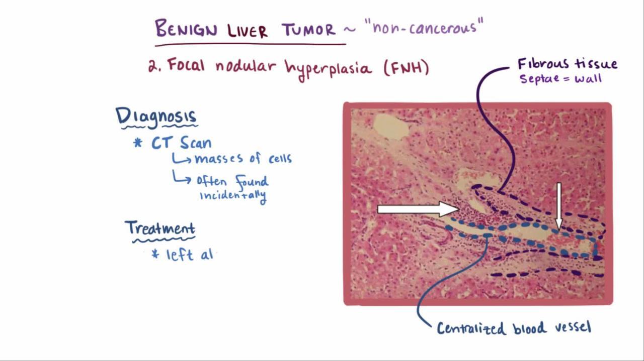
Focal Nodular Hyperplasia
This benign liver mass is the second most common solid liver lesion (1). Diagnosis is usually based on MRI or CT with hepatobiliary contrast; the classic appearance is a lesion with a central scar. But biopsy may be necessary for confirmation. Treatment is rarely needed unless symptomatic.
Focal nodular hyperplasia reference
1. Vilgrain V, Uzan F, Brancatelli G, Federle MP, Zappa M, Menu Y. Prevalence of hepatic hemangioma in patients with focal nodular hyperplasia: MR imaging analysis. Radiology. 2003;229(1):75-79. doi:10.1148/radiol.2291021284
Hemangiomas
Hemangiomas are usually small and asymptomatic; they occur in up to 20% of adults and more frequently in women (1). Symptoms are more likely if they are > 4 cm; symptoms include discomfort, fullness, and, less often, anorexia, nausea, early satiety, and pain secondary to bleeding or thrombosis. These tumors often have a characteristic highly vascular appearance. Hemangiomas are found incidentally during ultrasound, CT, or MRI. CT typically shows a well-demarcated, hypodense mass; when contrast is used, there is early peripheral enhancement, followed by later centrifugal enhancement. Biopsy is usually not necessary, and treatment is usually not indicated. Resection can be considered if symptoms are troublesome or if a hemangioma is rapidly enlarging.
In infants, hemangiomas often regress spontaneously. However, large hepatic hemangiomas occasionally cause arteriovenous shunting sufficient to cause heart failure and sometimes consumption coagulopathy. Treatment may include beta blockers, systemic corticosteroids and other immunomodulatory therapy, medical treatment of heart failure when necessary, surgical removal, selective hepatic artery embolization, and, rarely, liver transplantation (2).
References for hemangiomas
1. Frenette C, Mendiratta-Lala M, Salgia R, Wong RJ, Sauer BG, Pillai A . ACG Clinical Guideline: Focal Liver Lesions. Am J Gastroenterol. 2024;119(7):1235-1271. doi:10.14309/ajg.0000000000002857. doi:10.14309/ajg.0000000000002857
2. Northup PG, Garcia-Pagan JC, Garcia-Tsao G, et al. Vascular Liver Disorders, Portal Vein Thrombosis, and Procedural Bleeding in Patients With Liver Disease: 2020 Practice Guidance by the American Association for the Study of Liver Diseases. Hepatology. 2021;73(1):366-413. doi:10.1002/hep.31646
Other Benign Tumors
Lipomas (usually asymptomatic) and localized fibrous tumors (eg, fibromas) rarely occur in the liver.
Benign bile duct adenomas are rare, inconsequential, and usually detected incidentally. They are sometimes mistaken for metastatic cancer.
Drug Information for the Topic


