


Celiac disease is an immunologically mediated disease in genetically susceptible people caused by intolerance to gluten, resulting in mucosal inflammation and villous atrophy, which causes malabsorption. Symptoms usually include diarrhea and abdominal discomfort. Diagnosis is by small-bowel biopsies showing characteristic though not specific pathologic changes of villous atrophy that resolve with a strict gluten-free diet.


")

Celiac disease is a malabsorption disorder (1).
Reference
1. Stanciu D, Staykov H, Dragomanova S, et al. Gluten Unraveled: Latest Insights on Terminology, Diagnosis, Pathophysiology, Dietary Strategies, and Intestinal Microbiota Modulations-A Decade in Review. Nutrients. 2024;16(21):3636. Published 2024 Oct 25. doi:10.3390/nu16213636
Etiology of Celiac Disease
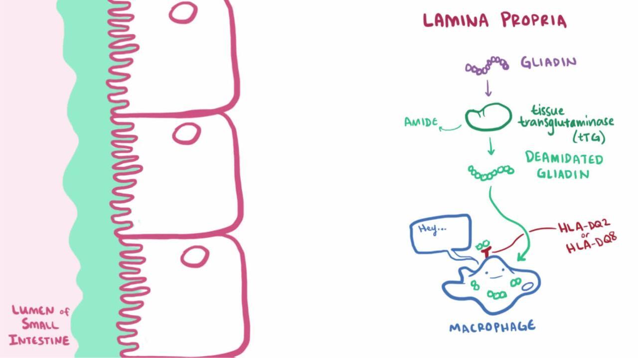
Celiac disease is a hereditary disorder caused by sensitivity to the gliadin fraction of gluten, a protein found in wheat; similar proteins are present in rye and barley. In a genetically susceptible person, gluten-sensitive T cells are activated when gluten-derived peptide epitopes are presented. The inflammatory response causes characteristic mucosal villous atrophy in the small bowel.

Epidemiology of Celiac Disease
Celiac disease may affect up to 1.4% of the global population, based on serologic screens of blood donors (1). The global prevalence of biopsy-proven celiac disease is about half that and varies widely from 0.4 to 0.8% depending on region.
The disease affects approximately 7.5% of first-degree relatives, with a higher prevalence in females than males (2). Onset is generally in childhood but may occur later.
Patients who have other diseases, such as lymphocytic colitis, Down syndrome, type 1 diabetes mellitus, and autoimmune (Hashimoto) thyroiditis, are at risk of developing celiac disease.
Epidemiology references
1. Singh P, Arora A, Strand TA, et al. Global Prevalence of Celiac Disease: Systematic Review and Meta-analysis. Clin Gastroenterol Hepatol. 2018;16(6):823-836.e2. doi:10.1016/j.cgh.2017.06.037
2. Singh P, Arora S, Lal S, Strand TA, Makharia GK. Risk of Celiac Disease in the First- and Second-Degree Relatives of Patients With Celiac Disease: A Systematic Review and Meta-Analysis. Am J Gastroenterol. 2015;110(11):1539-1548. doi:10.1038/ajg.2015.296
Symptoms and Signs of Celiac Disease
The clinical presentation varies enough that no typical presentation exists. Some patients are asymptomatic or have only signs of nutritional deficiency. Others have significant gastrointestinal symptoms.
Celiac disease can manifest in infancy and childhood after introduction of cereals into the diet. The child has failure to thrive, apathy, anorexia, pallor, generalized hypotonia, abdominal distention, and muscle wasting. Stools are soft, bulky, clay-colored, and foul-smelling. Older children may present with anemia or failure to grow normally.
In adults, lassitude, weakness, and anorexia are most common. Mild and intermittent diarrhea is sometimes the presenting symptom. Steatorrhea (foul-smelling, pale, bulky, and greasy stools) ranges from mild to severe (7 to 50 g of fat/day). Some patients have weight loss, rarely enough to become underweight. Anemia, glossitis, angular stomatitis, and aphthous ulcers are usually seen in these patients. Manifestations of vitamin D and calcium deficiencies (eg, osteomalacia, osteopenia, osteoporosis) are common. Both men and women may have reduced fertility; women may not have menstrual periods.In adults, lassitude, weakness, and anorexia are most common. Mild and intermittent diarrhea is sometimes the presenting symptom. Steatorrhea (foul-smelling, pale, bulky, and greasy stools) ranges from mild to severe (7 to 50 g of fat/day). Some patients have weight loss, rarely enough to become underweight. Anemia, glossitis, angular stomatitis, and aphthous ulcers are usually seen in these patients. Manifestations of vitamin D and calcium deficiencies (eg, osteomalacia, osteopenia, osteoporosis) are common. Both men and women may have reduced fertility; women may not have menstrual periods.
Approximately 17% of patients have dermatitis herpetiformis, an intensely pruritic papulovesicular rash that is symmetrically distributed over the extensor areas of the elbows, knees, buttocks, shoulders, and scalp (1). This rash can be induced by a high-gluten diet.


Symptoms and signs reference
1. Reunala T, Salmi TT, Hervonen K, Kaukinen K, Collin P. Dermatitis Herpetiformis: A Common Extraintestinal Manifestation of Coeliac Disease. Nutrients. 2018;10(5):602. Published 2018 May 12. doi:10.3390/nu10050602
Diagnosis of Celiac Disease
Serologic markers
Small-bowel biopsy
(See also the American College of Gastroenterology's 2023 Guidelines Update: Diagnosis and Management of Celiac Disease.)
The diagnosis of celiac disease is suspected clinically and by laboratory abnormalities suggestive of malabsorption. Family incidence is a valuable clue. Celiac disease should be strongly considered in a patient with iron deficiency without obvious gastrointestinal bleeding.
Confirmation requires a small-bowel biopsy from the second portion of the duodenum. Findings include lack or shortening of villi (villous atrophy), increased intraepithelial cells, and crypt hyperplasia. However, such findings can also occur in tropical sprue, severe small intestinal bacterial overgrowth, eosinophilic enteritis, infectious enteritis (eg, giardiasis), and lymphoma.


Top image: BIOPHOTO ASSOCIATES/SCIENCE PHOTO LIBRARY
Bottom image: INNERSPACE IMAGING/SCIENCE PHOTO LIBRARY
Because biopsy lacks specificity, serologic markers can aid diagnosis. Tissue transglutaminase antibody (tTG)-IgA antibody and endomysial IgA antibody (EMA—an antibody against an intestinal connective tissue protein) have sensitivity and specificity > 90% (1). These markers can also be used to screen populations with high prevalence of celiac disease, including first-degree relatives of affected patients and patients with diseases that occur at a greater frequency in association with celiac disease. If either test is positive, the patient should have a diagnostic small-bowel biopsy. If both are negative, celiac disease is extremely unlikely. These antibodies decrease in titer in patients on a gluten-free diet and thus are useful in monitoring dietary adherence. All diagnostic serologic testing should be performed with patients following a gluten-containing diet.
Serum IgA levels should be measured concurrently with tTG testing because IgA deficiency can lead to false-negative results. If IgA deficiency is detected, testing for IgG antibodies against tTG and deaminated gliadin peptide (DGP) is recommended.
Histocompatibility testing can be useful in selected clinical situations. More than 95% of celiac patients have the human leukocyte antigen (HLA)-DQ2 or HLA-DQ8 haplotype (2), although these haplotypes are not particularly specific for celiac disease. However, given the high sensitivity, testing that fails to show HLA-DQ2 or -DQ8 can effectively rule out celiac disease when biopsy and serologic markers are not concordant.
Other laboratory abnormalities often occur and should be sought. They include anemia (iron-deficiency anemia in children and folate-deficiency anemia in adults); hypoalbuminemia, hypocalcemia, hypokalemia, hyponatremia, elevated alkaline phosphatase, and prolonged prothrombin time.
Malabsorption tests are not specific for celiac disease. If these tests are performed, common findings include steatorrhea of 10 to 40 g/day and abnormal results with D-xylose and (in severe ileal disease) positive Schilling tests.
Nonceliac gluten sensitivity (NCGS), or gluten intolerance, is a nonimmune–mediated reaction to the ingestion of gluten. Similar gastrointestinal symptoms are noted as in patients with celiac disease, but biopsies will show normal villi and serologic markers will exclude celiac disease (and wheat allergy). There are some reports of nonspecific serologic markers that may be elevated in NCGS (3). As compared to celiac disease, gluten sensitivity has no serious negative effects on overall health, and its effects are mainly limited to uncomfortable gastrointestinal symptoms. As with celiac disease, the treatment is gluten avoidance.
Pearls & Pitfalls
|
Diagnosis references
1. Rubio-Tapia A, Hill ID, Semrad C, et al. American College of Gastroenterology Guidelines Update: Diagnosis and Management of Celiac Disease [published correction appears in Am J Gastroenterol. 2024 Jul 1;119(7):1441. doi: 10.14309/ajg.0000000000002210]. Am J Gastroenterol. 2023;118(1):59-76. doi:10.14309/ajg.00000000000020751. Epub 2022 Sep 21. Erratum in: Am J Gastroenterol. 2024 Jul 1;119(7):1441. doi: 10.14309/ajg.0000000000002210
2. Kaukinen K, Partanen J, Mäki M, Collin P. HLA-DQ typing in the diagnosis of celiac disease. Am J Gastroenterol. 97(3):695–699, 2002. doi: 10.1111/j.1572-0241.2002.05471.x
3. Hill ID, Fasano A, Guandalini S, et al. NASPGHAN Clinical Report on the Diagnosis and Treatment of Gluten-related Disorders. J Pediatr Gastroenterol Nutr. 2016;63(1):156-165. doi:10.1097/MPG.0000000000001216
Treatment of Celiac Disease
Gluten-free diet
Supplements to replace any serious deficiencies
(See also the American College of Gastroenterology's 2023 Guidelines Update: Diagnosis and Management of Celiac Disease.)
Treatment of celiac disease is a gluten-free diet (avoiding foods containing wheat, rye, or barley). Gluten is widely used in commercially prepared foods, so patients need a detailed list of foods to avoid. Patients are encouraged to consult a dietitian and join a celiac support group as well (English-language groups include Beyond Celiac or the Celiac Disease Foundation). The response to a gluten-free diet is usually rapid, and symptoms resolve in 1 to 2 weeks. Ingesting even small amounts of food containing gluten, however, may prevent remission or induce relapse.
Small-bowel biopsy should be repeated after 3 to 6 months of a gluten-free diet. If abnormalities persist, other causes of villous atrophy (eg, lymphoma) should be considered. Lessening of symptoms and improvement in small-bowel morphology are accompanied by a decrease in anti-tissue transglutaminase antibody and anti-endomysial antibody titers.
Supplementary vitamins, minerals, and hematinics may be given, depending on the deficiencies. Mild cases may not require supplementation, whereas severe cases may require comprehensive replacement. For adults, replacement includes oral ferrous sulfate, oral folate, calcium supplements, and any standard multivitamin. Sometimes children (but rarely adults) who are seriously ill on initial diagnosis require bowel rest and total parenteral nutrition.
If a patient responds poorly to gluten withdrawal, either the diagnosis is incorrect or the disease has become refractory. Corticosteroids can control symptoms in refractory disease.
Prognosis for Celiac Disease
Complications of celiac disease include refractory disease, collagenous sprue, and intestinal lymphomas.
Intestinal lymphomas affect 6 to 8% of patients with celiac disease, usually manifesting after 20 to 40 years of disease. The incidence of other gastrointestinal cancers (eg, carcinoma of the esophagus or oropharynx, small-bowel adenocarcinoma) also increases (1). Adherence to a gluten-free diet can significantly reduce the risk of cancer.
If people who have been doing well on a gluten-free diet for a long time once again develop symptoms of celiac disease, physicians usually do upper endoscopy with small-bowel biopsy and/or capsule endoscopy to check for signs of intestinal lymphoma.
Prognosis reference
1. Ilus T, Kaukinen K, Virta LJ, et al. Incidence of malignancies in diagnosed celiac patients: A population-based estimate. Am J Gastroenterol. 109(9):1471–1477, 2014. doi: 10.1038/ajg.2014.194
Key Points
Celiac disease involves an inflammatory response to gluten that causes villous atrophy and malabsorption.
Prevalence varies in different populations worldwide.
Suspect the diagnosis if the serologic markers anti-tissue transglutaminase antibody and anti-endomysial antibody are present, and confirm the diagnosis with a small-bowel biopsy.
Instruct the patient to follow a gluten-free diet and replace any vitamin or mineral deficiencies.


