


Peritonsillar abscess requires incision and drainage or needle aspiration.
Topic Resources


Peritonsillar abscess must be distinguished from peritonsillar cellulitis (see Peritonsillar Abscess and Cellulitis) and from parapharyngeal abscess, a deep neck abscess. Cellulitis does not require drainage, however, a parapharyngeal abscess should be drained as an operative procedure.
There are 2 methods for draining a peritonsillar abscess: needle aspiration or incision and drainage. Needle aspiration is cost-effective, less invasive and can be performed under local anesthesia. Incision and drainage is more invasive, but may have a lower recurrence rate. The choice of method depends largely on the clinician's experience and the severity of the abscess, as there is insufficient evidence to recommend one approach over the other (1).
Indications for Draining a Peritonsillar Abscess
Clinically apparent peritonsillar abscess: Incision and drainage or needle aspiration
Possible peritonsillar abscess: Needle aspiration for diagnosis and treatment
The presence of significant pain, trismus, dysphagia, respiratory distress, or the lack of response to antibiotic therapy should warrant drainage of a peritonsillar abscess (2).
If the diagnosis is uncertain, point-of-care ultrasound or needle aspiration can be done to confirm the presence of an abscess. Alternatives include CT scan or, for mildly ill patients, discharge on antibiotics with close follow-up. See Peritonsillar Abscess and Cellulitis for further information.
Contraindications to Draining a Peritonsillar Abscess
Absolute contraindications
Intractable trismus
Relative contraindications
Poor patient cooperation
Coagulopathy
Uncertain diagnosis (for incision and drainage)
Complications of Draining a Peritonsillar Abscess
Aspiration of blood
Hemorrhage
Puncture of the carotid artery
Incomplete drainage of the abscess
Equipment for Draining a Peritonsillar Abscess
Gloves
Protective eyewear
Mask
Gown
Medications for IV analgesia and sedation
Local anesthetic (eg, 1% lidocaine with epinephrine), 25- and 20- to 22-gauge needles, and 5-mL syringeLocal anesthetic (eg, 1% lidocaine with epinephrine), 25- and 20- to 22-gauge needles, and 5-mL syringe
Topical anesthetic spray (eg, 4% lidocaine)Topical anesthetic spray (eg, 4% lidocaine)
Tongue depressor
Headlamp
Frazier-tip or Yankauer suction catheter attached to wall suction
For aspiration, a 10-mL syringe with 18- or 20-gauge needle
For incision and drainage, a scalpel with a No. 11 or 15 blade
For incision and drainage, a tonsil clamp
Normal saline or peroxide-saline solution
Additional Considerations for Draining a Peritonsillar Abscess
Needle aspiration may miss the abscess cavity and result in misdiagnosis as peritonsillar cellulitis. Thus, if an abscess is still suspected (eg, based on clinical or imaging findings), some clinicians treat patients with IV antibiotics, corticosteroids, and close observation, sometimes as an inpatient, even if needle aspiration yields no pus (3).

Relevant Anatomy for Draining a Peritonsillar Abscess
The tonsils are located between the anterior and posterior pillars of the throat. The lateral wall of the tonsil is adjacent to the superior pharyngeal constrictor muscle.
A peritonsillar abscess is located between the tonsil capsule, the superior pharyngeal constrictor muscle, and the palatopharyngeus muscle. The abscess is not within the tonsil.
The internal carotid artery lies about 2.5 cm posterolateral to the tonsil.

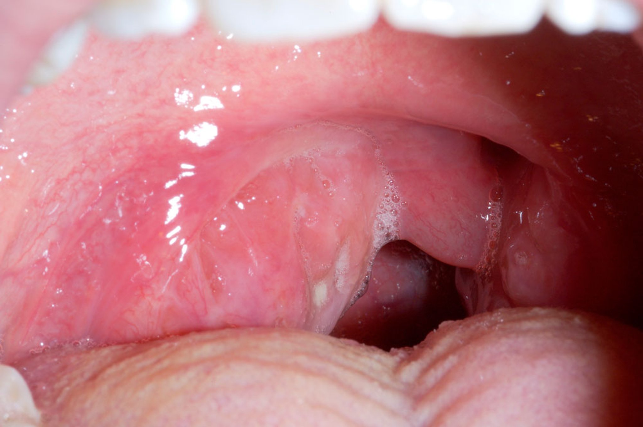
DR P. MARAZZI/SCIENCE PHOTO LIBRARY
Positioning for Draining a Peritonsillar Abscess
Patient should sit upright with a support behind the head to prevent sudden backward movement.
Step-by-Step Description of Draining a Peritonsillar Abscess
Consider whether IV analgesia is necessary (it is usually not if an adequate explanation and local anesthesia are given). If needed, give fentanyl 1 to 3 mcg/kg, titrated if necessary, a few minutes before the procedure.Consider whether IV analgesia is necessary (it is usually not if an adequate explanation and local anesthesia are given). If needed, give fentanyl 1 to 3 mcg/kg, titrated if necessary, a few minutes before the procedure.
Spray the topical anesthetic and wait several minutes for it to take effect.
Have an assistant retract the cheek laterally to improve visibility.
Push the tongue out of the way using a tongue depressor or finger.
Identify the most prominent part of the abscess. Point-of-care ultrasound is sometimes used to localize the abscess.
Inject 2 to 3 mL of anesthetic (1% lidocaine with epinephrine) into the mucosa using a 25-gauge needle attached to the 5-mL syringe. Inject 2 to 3 mL of anesthetic (1% lidocaine with epinephrine) into the mucosa using a 25-gauge needle attached to the 5-mL syringe.
Some clinicians give a dose of IV corticosteroids (eg, dexamethasone 10 mg, methylprednisolone 60 mg) to decrease symptoms.Some clinicians give a dose of IV corticosteroids (eg, dexamethasone 10 mg, methylprednisolone 60 mg) to decrease symptoms.
For needle aspiration
Use the 10-mL syringe with an 18- or 20-gauge needle
Apply continuous suction and direct the needle in the sagittal plane (anterior to posterior) and not to the side (laterally). This is important to avoid the carotid artery. This may be done with ultrasound guidance.
Aspirate the most prominent area first; it is usually the superior pole. If no pus is aspirated, aspirate the middle, then inferior pole. Do not aspirate the tonsil itself.
Typically 2 to 6 mL of pus is obtained. Send a sample for culture.
For incision and drainage
Warn the patient that pus will flow and must be spit out.
Use a scalpel with a No. 15 blade or a No. 11 with tape covering all but 0.5 to 1.0 cm of the blade.
Make an 0.5 cm incision in the anterior-to-posterior direction over the most prominent (or alternatively, the most fluctuant) area, or the location where needle aspiration (if done initially) identified pus.
Use a suction catheter to remove pus and blood. Some bleeding is expected after the incision.
Place a closed tonsil clamp into the incised opening and gently open it to break up any loculations; then remove the clamp.
Finally, have the patient rinse and gargle with a saline or a dilute peroxide-saline solution.
Aftercare for Draining a Peritonsillar Abscess
Observe the patient for 1 hour after the procedure for complications such as bleeding, and to ensure that the patient can tolerate fluids.
Discharge on oral antibiotics and warm saline rinses. The patient must be advised to follow up in 24 hours.
Patients with excessive bleeding, aspiration, or who are unable to take oral antibiotics require prolonged observation or hospitalization.
Patients who have had multiple abscesses should usually have elective tonsillectomy after 4 to 6 weeks to prevent abscess recurrence.
Antibiotics should be continued for 10 days. Examples of appropriate empiric antibiotics are penicillin, 1st-generation cephalosporins, and clindamycin. Preferably, culture-directed antibiotics are then prescribed. If methicillin-resistant Antibiotics should be continued for 10 days. Examples of appropriate empiric antibiotics are penicillin, 1st-generation cephalosporins, and clindamycin. Preferably, culture-directed antibiotics are then prescribed. If methicillin-resistantStaphylococcus aureus (MRSA) is a possibility, empiric antibiotics should be broadened to cover this.
Warnings and Common Errors for Draining a Peritonsillar Abscess
Oversedating the patient and risking aspiration
Injecting anesthetic directly into the abscess cavity (because this is painful)
Inserting the needle or scalpel blade too deeply (because this risks penetrating the carotid artery); if pus is not obtained at 1-cm depth, do not go deeper.
For needle aspiration, not ensuring that the needle is inserted in the sagittal plane (anterior to posterior). Do not insert the needle to the side (laterally) in the direction of the carotid artery.
Tips and Tricks for Draining a Peritonsillar Abscess
A headlamp is essential because it allows the use of both hands: one to perform the needle aspiration and the other to depress the tongue with a tongue blade.
When anesthetic is injected at the correct depth, the mucosa should blanch because of epinephrine-induced vasoconstriction.When anesthetic is injected at the correct depth, the mucosa should blanch because of epinephrine-induced vasoconstriction.
For needle aspiration, to limit depth of penetration, some clinicians cut off the distal 1 cm of the plastic needle sheath and replace it over the needle, thus leaving only 1 cm of needle protruding. Tape the sheath onto the syringe so it does not fall off and become aspirated.
Similarly for incision and drainage, some clinicians apply tape to all but the distal 0.5 to 1 cm of the scalpel blade as a depth guide.
If pus continues to drain from the needle puncture site, repeat aspiration or incision and drainage may be indicated.
References
1. Chang BA, Thamboo A, Burton MJ, Diamond C, et al. Needle aspiration versus incision and drainage for the treatment of peritonsillar abscess. Cochrane Database Syst Rev. 2016;12(12):CD006287. Published 2016 Dec 23. doi:10.1002/14651858.CD006287.pub4
2. Galioto NJ. Peritonsillar Abscess. Am Fam Physician. 2017;95(8):501-506.
3. Powell J, Wilson JA. An evidence-based review of peritonsillar abscess. Clin Otolaryngol. 2012;37(2):136-145. doi:10.1111/j.1749-4486.2012.02452.x


