


- Overview of Arrhythmogenic Cardiomyopathies
- Arrhythmogenic Right Ventricular Cardiomyopathy
- Lamin A/C Cardiomyopathies (Cardiolaminopathies)
- Non-Compaction Cardiomyopathy
- Cardiac Sarcoidosis
- Hypertrophic Cardiomyopathy Arrhythmias
- Overview of Channelopathies
- Brugada Syndrome
- Catecholaminergic Polymorphic Ventricular Tachycardia
- Early Repolarization Syndrome
- Long QT Interval Syndromes
- Isolated Progressive Cardiac Conduction Disease
- Short QT Interval Syndromes
The long QT interval syndromes (LQTS) result from any congenital or acquired disorder of cardiac ion channel function or regulation (channelopathy) that prolongs ventricular myocyte action potential duration as reflected by prolongation of the rate-corrected QT interval on the ECG. Patients are at risk for torsades de pointes polymorphic ventricular tachycardia, which may cease spontaneously or degenerate into ventricular fibrillation. Diagnosis is by clinical criteria and ECG, sometimes with exercise and/or provocative testing. Treatment is avoidance of triggers, beta-blockade, and sometimes an implantable cardioverter-defibrillator.


*")

(See also Overview of Arrhythmias and Overview of Channelopathies.)
Long QT interval syndromes can be acquired, congenital, or both. (For discussion of acquired causes, see Torsades de Pointes Ventricular Tachycardia). The incidence of congenital LQTS is approximately 1 in 2000 (1) and is the cause of about 15% of cardiac arrests that are unexplained after clinical evaluation that includes and ECG, an echocardiogram, and ruling out coronary artery disease (2).
General references
1. Schwartz PJ, Stramba-Badiale M, Crotti L, et al: Prevalence of the congenital long-QT syndrome. Circulation 120(18):1761–1767, 2009. doi:10.1161/CIRCULATIONAHA.109.863209
2. Krahn AD, Healey JS, Chauhan V, et al: Systematic assessment of patients with unexplained cardiac arrest: Cardiac Arrest Survivors With Preserved Ejection Fraction Registry (CASPER). Circulation 120(4):278–285, 2009. doi: 10.1161/CIRCULATIONAHA.109.853143
Pathophysiology of Long QT Interval Syndromes
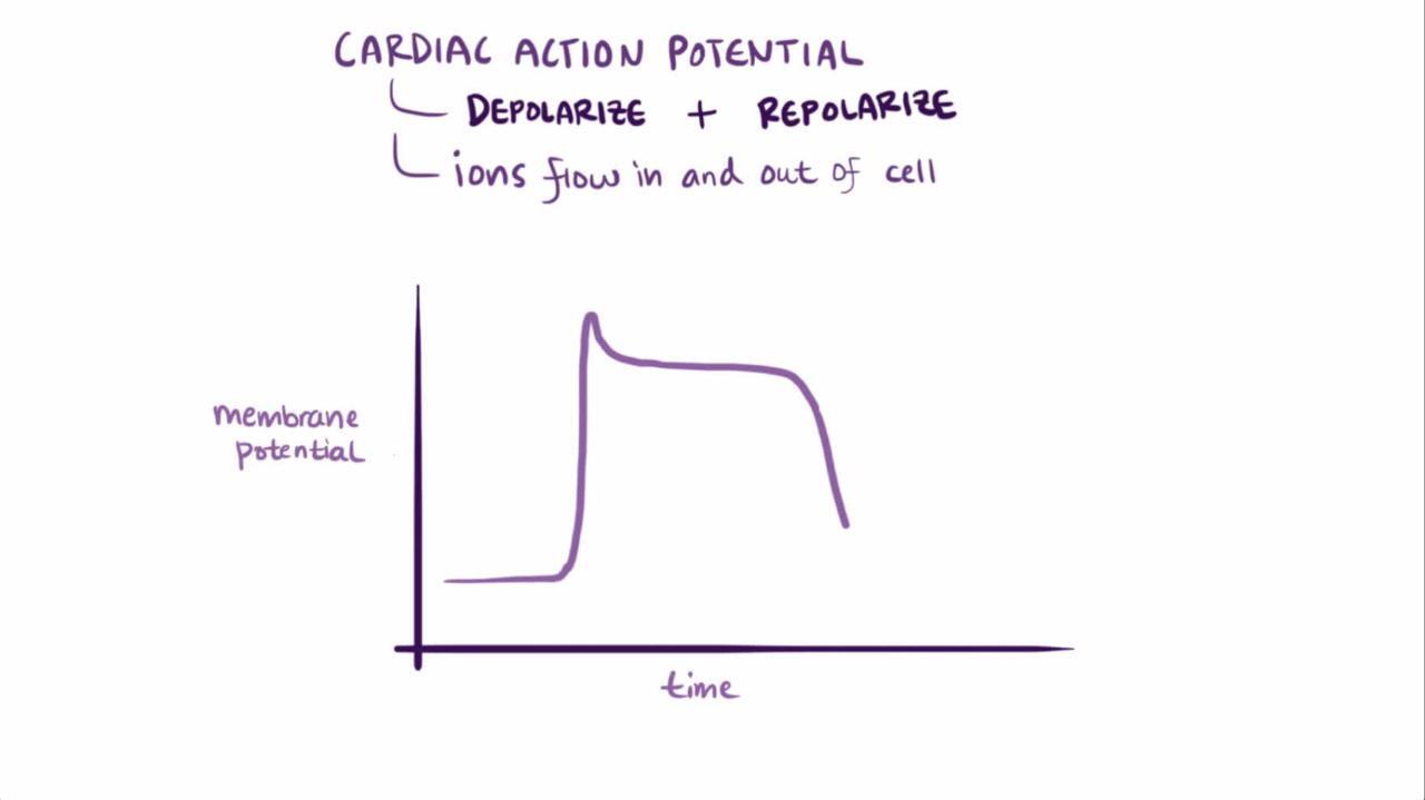
The congenital long QT interval syndromes result from genetic disorders of cardiac ion channel function or regulation (channelopathies) that prolong ventricular myocyte action potential duration as reflected by prolongation of the rate-corrected QT interval on the ECG (QTc, typically calculated using Bazett's formula).

The dysfunction may involve
Loss of function of repolarizing potassium current channels OR
Gain of function of depolarizing sodium or depolarizing calcium current channels
Prolongation of action potentials increases the probability of transmembrane voltage oscillations occurring during the depolarized myocyte action potential (early afterdepolarizations). If the action potential durations of myocytes in a local area vary, these oscillations may reactivate neighboring myocytes that have repolarized and thus create torsades de pointes ventricular tachycardia (TdeP VT). The risk of TdeP VT is dependent on the degree of QTc prolongation, particularly if it is > 0.50 second (1).
LQTS (particularly LQTS3) may also cause paroxysmal atrial fibrillation.
Predisposing factors for arrhythmia
The occurrence of TdeP VT is favored by any condition that further prolongs action potential duration, including female sex, bradycardia, hypokalemia, hypomagnesemia, and hypothyroidism. Other risk factors include slow or irregular ventricular rate, acute intracranial disasters (eg, bleeding, stroke, traumatic brain injury), eating disorders, organophosphate poisoning, and structural heart disease (especially acute ischemia, myocarditis, and ventricular hypertrophy). Many medications are risk factors, particularly class Ia, Ic, and III antiarrhythmic medications, as well as other medications, including tricyclic antidepressants, phenothiazines, and certain antivirals and antifungals (see CredibleMeds for up-to-date information). Often, several of these factors are present (2).
Pathophysiology references
1. Sauer AJ, Moss AJ, McNitt S, et al: Long QT syndrome in adults. J Am Coll Cardiol 49(3):329–337, 2007. doi: 10.1016/j.jacc.2006.08.057
2. Roden DM: Long QT syndrome: reduced repolarization reserve and the genetic link. J Intern Med 259(1):59–69, 2006. doi: 10.1111/j.1365-2796.2005.01589.x
Etiology of Long QT Interval Syndromes
Long QT interval syndromes are classified based on the specific gene that has mutated. However, a specific genetic abnormality is identified in only 70 to 85% of cases (1); likelihood of detecting an abnormality varies depending on clinical factors present.
More than 15 forms of LQTS have been described, but most cases fall into 3 subgroups:
Long QT syndrome type 1 (LQTS1): Loss-of-function mutation of gene KCNQ1, which encodes an adrenergic-sensitive Kv7.1 channel responsible for the slow outward potassium current (IKs)
Long QT syndrome type 2 (LQTS2): Loss-of-function mutation of gene KCNH2, which encodes the hERG channel responsible for the rapid outward potassium current (IKr)
Long QT syndrome type 3 (LQTS3): Gain-of-function mutation of gene SCN5A, which encodes the Nav1.5 channel responsible for the inward sodium current (INa).
The vast majority of cases are LQTS1, LQTS2, or LQTS3. These 3 forms are inherited as autosomal dominant disorders with incomplete penetrance.
Rare forms of LQTS with additional clinical features have been described including Jervell and Lange Nielsen syndrome (with congenital neural deafness), Andersen-Tawil syndrome (with period paralysis and craniofacial dysmorphisms), and Timothy syndrome (with craniofacial dysmorphisms, immunodeficiency, congenital heart disease, developmental delay, and syndactyly).

Etiology reference
1. Wilde AAM, Semsarian C, Márquez MF, et al: European Heart Rhythm Association (EHRA)/Heart Rhythm Society (HRS)/Asia Pacific Heart Rhythm Society (APHRS)/Latin American Heart Rhythm Society (LAHRS) Expert Consensus Statement on the state of genetic testing for cardiac diseases. J Arrhythm 38(4):491–553, 2022. doi: 10.1002/joa3.12717
Symptoms and Signs of Long QT Interval Syndromes
The LQTS are asymptomatic unless TdeP VT occurs, which can cause palpitations, near syncope, or syncope. Some patients experience myoclonic jerks during syncope; they may incorrectly have a diagnosis of epilepsy pursued. Because the ventricular action potential duration decreases with increasing heart rate, TdeP VT is often self-terminating. However, it may degenerate into ventricular fibrillation and cause cardiac arrest and sudden death. In patients with LQTS, there is a 5-year risk of a first episode of life-threatening arrhythmia ranging from approximately 0.3% to 17% (1). Predictors include the following
Age
Sex
Use of beta-blocker therapy (yes or no)
Syncope (none, while not on beta-blocker therapy, or while on beta-blocker therapy)
QTc interval
Genotype (if available; higher in LQTS2 and LQTS3 than in LQTS-1)
An online risk calculator for patients who have not yet had a life-threatening arrhythmia is available (1).
Symptoms and signs reference
1. Wang M, Peterson DR, Pagan E, et al: Assessment of absolute risk of life-threatening cardiac events in long QT syndrome patients. Front Cardiovasc Med 2022;9:988951. Published 2022 Oct 7. doi:10.3389/fcvm.2022.988951
Diagnosis of Long QT Interval Syndromes
Characteristic clinical and electrocardiographic manifestations
Sometimes exercise testing
Sometimes ambulatory ECG monitoring
Sometimes provocative testing using IV epinephrine or isoproterenol Sometimes provocative testing using IV epinephrine or isoproterenol
Often genetic testing
Screening of relatives
Diagnosis should be considered in patients with unexplained cardiac arrest or syncope or a family history of such when the affected people do not have structural heart disease. It should also be considered in people who are discovered to have a long QT interval when ECG is done for other reasons.
A long QT interval is diagnosed by ECG showing prolongation of the rate-corrected QT interval (QTc). Normal QTc values are < 0.43 second for males and < 0.45 second for females and are considered prolonged when > 0.45 second for males or > 0.47 second for females (1).
However, given the multiplicity of factors affecting the QTc, a normal QTc does not exclude the diagnosis. Nevertheless, at the moment of torsade de pointes VT, the QTc is essentially always prolonged.
When a patient has a significantly prolonged QT interval and documented torsade de pointes VT in the absence of other causes of a prolonged QT interval, the diagnosis of a congenital long QT interval syndrome is established. Patients with borderline QT intervals suspected of having LQTS should have exercise testing, because some abnormalities appear only during exercise. Ambulatory ECG monitoring may also disclose transient ventricular repolarization abnormalities. In patients with a normal QTc interval, provocative testing with IV isoproterenol or epinephrine may disclose a concealed long QTc and should be considered in patients with an intermediate probability of a congenital LQTS. in the absence of other causes of a prolonged QT interval, the diagnosis of a congenital long QT interval syndrome is established. Patients with borderline QT intervals suspected of having LQTS should have exercise testing, because some abnormalities appear only during exercise. Ambulatory ECG monitoring may also disclose transient ventricular repolarization abnormalities. In patients with a normal QTc interval, provocative testing with IV isoproterenol or epinephrine may disclose a concealed long QTc and should be considered in patients with an intermediate probability of a congenital LQTS.
Because not all patients with a long QT interval have congenital long QT syndrome and because not all patients with a congenital long QT syndrome have a long QT interval on any given ECG, the Schwartz score has been developed to estimate the probability of a congenital LQTS (see table Schwartz Score for Long QT Syndrome). Probability is estimated as low, intermediate, or high based on clinical, ECG, and exercise testing criteria, provided that the patient is not presently exposed to any environmental causes of QT-interval prolongation. The score can be used to establish candidacy for genetic testing, which can be time-consuming and expensive because of the multiple gene variants to be tested for. Patients with a low probability of a congenital LQTS do not need genetic testing, but patients in whom the probability is intermediate or high do. High-probability patients without a detected genetic abnormality may be considered to represent one of the 15 to 30% of patients with an unidentified mutation. Intermediate-probability patients who are gene mutation–negative are followed closely with repeated electrocardiographic examinations including ECG, ambulatory cardiac monitoring, and exercise testing (2, 3).
Schwartz Score for Long QT Syndrome (LQTS)*
CRITERIA | POINTS |
|---|---|
Patient History | |
Syncope with stress† | 2 |
Syncope without stress† | 1 |
Congenital deafness | 0.5 |
Family History | |
Family member with known LQTS‡ | 1 |
Family member with unexplained cardiac death before age 30 ‡ | 0.5 |
Electrocardiography§ | |
QTc ≥ 480 msec | 3 |
QTc 460-479 msec | 2 |
QTc 450-459 msec (males only) | 1 |
QTc ≥ 480 msec during 4th minute of recovery from an exercise test | 1 |
Torsades de pointe arrhythmia§ | 2 |
T wave alternans | 1 |
Notched T wave in 3 leads | 1 |
Resting heart rate < 2nd percentile for age | 0.5 |
* SCORE: ≤ 1 low probability; 1.5–3 intermediate probability; ≥ 3.5 high probability | |
† Mutually exclusive (ie, if syncope occurs with stress, no points are given for syncope without stress) | |
‡ Mutually exclusive | |
§ If torsades de pointe present, do not score syncope | |
Data from Schwartz PJ, Crotti L: QTc behavior during exercise and genetic testing for the long-QT syndrome. Circulation 124:2181–2184, 2011. doi: 10.1161/CIRCULATIONAHA.111.062182 | |
First-degree family members of the index case should have clinical evaluation (ie, to detect symptoms suggestive of arrhythmia) and ECG. Thereafter, the first-degree family members of any newly identified patients undergo similar assessment (cascade screening). Genetic testing is done when the index case has a known mutation. Exercise testing is done when the results could alter the Schwartz score probability result.
Some forms of LQTS are more associated with certain triggers than others.
LQTS1: Physical stress, particularly swimming, or emotional stress
LQTS2: Sudden loud noises like an alarm clock
LQTS3: Sleep
Some forms of LQTS are also associated with particular ECG patterns
LQTS1: Wide T waves
LQTS2: Low voltage, notched T-waves
LQTS3: Long-ST segment with normal appearing T-waves
However, neither the triggers nor the ECG findings are very specific and should not be used to confirm type or to direct genetic testing.
Diagnosis references
1. Goldenberg I, Moss AJ, Zareba W: QT interval: how to measure it and what is "normal". J Cardiovasc Electrophysiol 2006 Mar;17(3):333-6. doi: 10.1111/j.1540-8167.2006.00408.x
2. Schwartz PJ, Moss AJ, Vincent GM, Crampton RS: Diagnostic criteria for the long QT syndrome: an update. Circulation 88:782–784, 1993. doi: 10.1161/01.cir.88.2.782
3. Schwartz PJ, Crotti L: QTc behavior during exercise and genetic testing for the long-QT syndrome. Circulation 124:2181–2184, 2011. doi: 10.1161/CIRCULATIONAHA.111.062182
Treatment of Long QT Interval Syndromes
Treatment of any VT/VF
Alleviation of predisposing causes and triggers, especially electrolyte abnormalities and use of certain medications
Usually beta-blockade
Sometimes mexiletineSometimes mexiletine
Sometimes an ICD
Sometimes left cardiac sympathetic denervation
Details of treatment of torsade de pointes ventricular tachycardia are discussed elsewhere but include cardioversion for torsade-induced VF and magnesium sulfate IV. Patients with frequent or long runs of torsades de pointes ventricular tachycardia may benefit from treatment to shorten the QT interval by increasing the heart rate using temporary pacing. Unlike medication-induced acquired torsade de pointes, the mainstay of acute IV pharmacotherapy is IV beta-blocker rather than IV isoproterenol.are discussed elsewhere but include cardioversion for torsade-induced VF and magnesium sulfate IV. Patients with frequent or long runs of torsades de pointes ventricular tachycardia may benefit from treatment to shorten the QT interval by increasing the heart rate using temporary pacing. Unlike medication-induced acquired torsade de pointes, the mainstay of acute IV pharmacotherapy is IV beta-blocker rather than IV isoproterenol.
Long-term treatment to prevent sudden death includes avoidance of specific triggers (including strenuous exercise in LQTS1 and LQTS2) and QTc-prolonging conditions. When possible, clinicians should stop any predisposing medications and prescribe alternatives (see CredibleMeds for up-to-date information). Patients, particularly those who will not accept exercise restrictions, should be counseled on the need for appropriate cautions (eg, availability of an automated external defibrillator during training and competition).
Beta-blocker therapy is recommended for most patients who have LQTS with QT interval prolongation or who are symptomatic (most effective with LQTS1 or LQTS2). When a beta-blocker is used, a long-acting beta-blocker without intrinsic sympathomimetic activity (eg, nadolol, slow-release propranolol) is preferred. When beta-blocker therapy is ineffective at relieving symptoms, mexiletine is added (most effective in LQTS3).Beta-blocker therapy is recommended for most patients who have LQTS with QT interval prolongation or who are symptomatic (most effective with LQTS1 or LQTS2). When a beta-blocker is used, a long-acting beta-blocker without intrinsic sympathomimetic activity (eg, nadolol, slow-release propranolol) is preferred. When beta-blocker therapy is ineffective at relieving symptoms, mexiletine is added (most effective in LQTS3).
Permanent pacing to increase the basal ventricular rate and to prevent post-extrasystolic pauses may reduce the probability of recurrent TdeP VT. An implantable cardioverter-defibrillator (ICD) is indicated in patients who have been resuscitated after cardiac arrest and in those who have cardiac syncope despite therapy with a beta-blocker (see table Indications for Implantable Cardioverter-Defibrillators in Ventricular Tachycardia and Ventricular Fibrillation).
Left heart (stellate ganglion) denervation may also be used (1).
Treatment reference
1. Savastano S, Schwartz PJ: Blocking nerves and saving lives: Left stellate ganglion block for electrical storms. Heart Rhythm 20(7):1039–1047, 2023. doi: 10.1016/j.hrthm.2022.11.025
Key Points
Congenital long QT interval syndromes may cause torsades de pointes ventricular tachycardia, ventricular fibrillation, and sudden death.
Numerous factors, particularly use of certain medications, increase the risk of ventricular arrhythmias.
Diagnosis is based on a combination of clinical and electrocardiographic criteria, including exercise and sometimes provocative testing.
Genetic testing is done on selected patients.
Family members should be screened.
Long-term management includes avoidance of triggers, use of beta-blockers, mexiletine, permanent pacing, an implantable cardioverter-defibrillator, or stellate ganglion denervation.Long-term management includes avoidance of triggers, use of beta-blockers, mexiletine, permanent pacing, an implantable cardioverter-defibrillator, or stellate ganglion denervation.


