


Almost half of all people in the United States report sleep-related problems. Disordered sleep can cause emotional disturbance, memory difficulty, poor motor skills, decreased work efficiency, and increased risk of traffic accidents. It can even contribute to cardiovascular disorders and mortality.
The most commonly reported sleep-related symptoms are insomnia and excessive daytime sleepiness (EDS).
Insomnia is difficulty falling or staying asleep, early awakening, or a sensation of unrefreshing sleep.
EDS is the tendency to fall asleep during normal waking hours.
EDS is not a disorder but a symptom of various sleep-related disorders. Insomnia can be a disorder, even if it exists in the context of other disorders, or can be a symptom of other disorders.
Parasomnias are abnormal sleep-related events (eg, night terrors, sleepwalking).
(See also Sleep Apnea and Sleep Problems in Children.)
Pathophysiology of Sleep
There are 2 states of sleep, each marked by characteristic physiologic changes:
Nonrapid eye movement (NREM): NREM sleep constitutes about 75 to 80% of total sleep time in adults. Heart rate and body temperature tend to decrease. NREM sleep consists of 3 stages (N1 to N3) in increasing depth of sleep. Slow, rolling eye movements, which characterize quiet wakefulness and early stage N1 sleep, disappear in deeper sleep stages. Muscle activity also decreases. Stage N2 sleep is characterized by K complexes and sleep spindles on the EEG (see figure Nonrapid Eye Movement [NREM] EEG). Stage N3 is referred to as deep sleep because arousal threshold is high; people may perceive this stage as high-quality sleep.
Rapid eye movement (REM): REM sleep follows each cycle of NREM sleep. It is characterized by low-voltage fast activity on the EEG and postural muscle atonia. Respiration rate and depth fluctuate dramatically. Most dreams occur during REM sleep. Normally, 20 to 25% of sleep is REM.
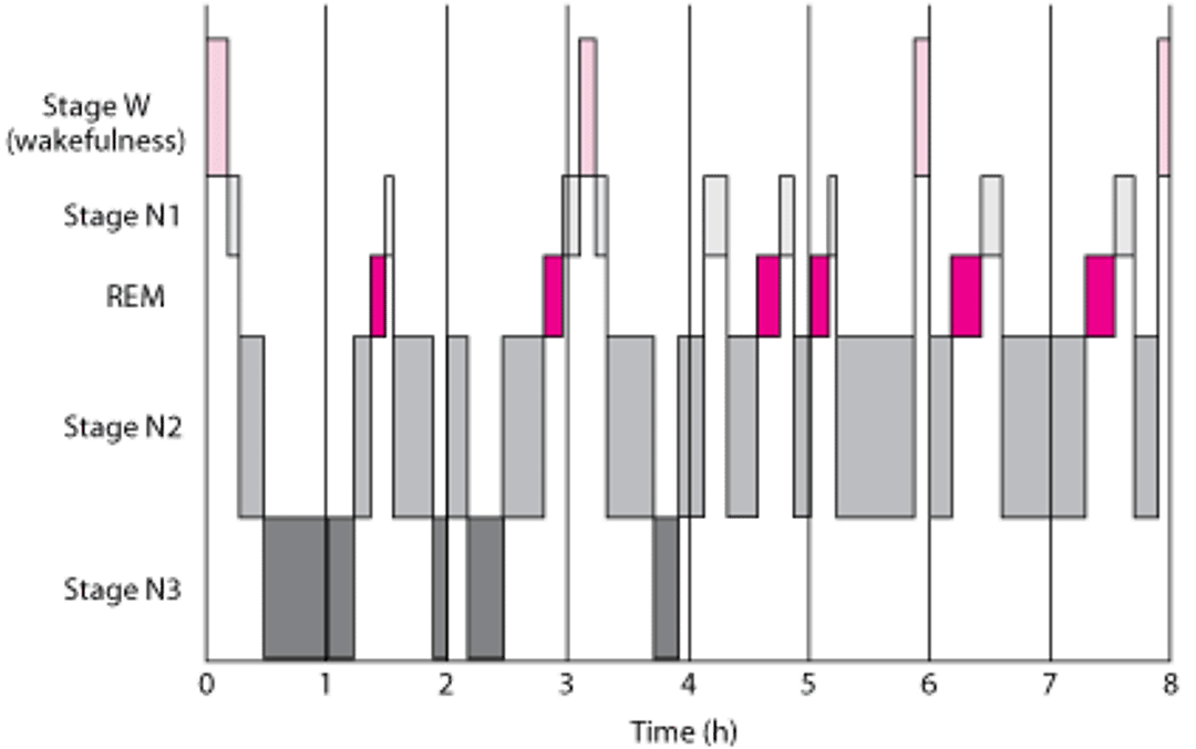
Progression through the 3 stages, typically followed by a brief interval of REM sleep, occurs cyclically 5 to 6 times a night (see figure Typical Sleep Pattern in Young Adults). Brief periods of wakefulness (stage W) occur periodically.
Nonrapid Eye Movement (NREM) EEG
These EEG tracings show characteristic theta waves, sleep spindles, and K complexes during stages 1 (N1), 2 (N2), and 3 (N3) NREM sleep. |


Rapid Eye Movement (REM) EEG
This figure includes an EEG tracing (showing characteristic sawtooth waves) and an eye tracing (showing rapid eye movements), which occur during REM sleep. In the bottom panel, the arrows represent sharply peaked conjugate eye movements from the right and left eyes during REM sleep. |

Individual sleep requirements vary widely, ranging from 6 to 10 hours/24 hours. Infants sleep a large part of the day; with aging, total sleep time and deep sleep (stage N3) tend to decrease, and sleep becomes more interrupted. In older people, stage N3 may disappear. These changes may account for increasing EDS and fatigue with aging, but their clinical significance is unclear.
Typical Sleep Pattern in Young Adults
Rapid eye movement (REM) sleep occurs cyclically throughout the night every 90–120 min. Brief periods of wakefulness (stage W) occur periodically. Sleep time is spent as follows:
|

Etiology of Sleep or Wakefulness Disorders
Some disorders can cause either insomnia or EDS (and sometimes both), and some cause only one or the other (see table Some Causes of Insomnia and Excessive Daytime Sleepiness).
Insomnia is most often caused by
An insomnia disorder (eg, adjustment sleep disorder, psychophysiologic insomnia)
Inadequate sleep hygiene
Psychiatric disorders, particularly mood, anxiety, and substance use disorders
Miscellaneous medical disorders such as cardiopulmonary disorders, musculoskeletal conditions, and chronic pain
EDS is most often caused by
Miscellaneous medical, neurologic (eg, narcolepsy, periodic limb movement disorder, and psychiatric disorders)
Circadian rhythm sleep disorders such as jet lag and shift work sleep disorders
Inadequate sleep hygiene refers to behaviors that are not conducive to sleep. They include
Consumption of caffeine or sympathomimetic or other stimulant drugs (typically near bedtime, but even in the afternoon for people who are particularly sensitive)
Exercise or excitement (eg, a thrilling television show, sports event) late in the evening
An irregular sleep-wake schedule
Patients who compensate for lost sleep by sleeping late or by napping may further fragment their nocturnal sleep.
Adjustment insomnia results from acute emotional stressors (eg, job loss, hospitalization, loss of a family member) that disrupt sleep.
Psychophysiologic insomnia is insomnia (regardless of cause) that persists well beyond resolution of precipitating factors, usually because patients feel anticipatory anxiety about the prospect of another sleepless night followed by another day of fatigue. Typically, patients spend hours in bed focusing on and brooding about their sleeplessness, and they have greater difficulty falling asleep in their own bedroom than falling asleep away from home.
Physical disorders that cause pain or discomfort (eg, arthritis, cancer, herniated disks), particularly those that worsen with movement, can cause transient awakenings and poor sleep quality. Nocturnal seizures can also interfere with sleep.
Most major mental disorders are associated with EDS and insomnia. About 80% of patients with major depression report EDS and insomnia; conversely, 40% of patients with chronic insomnia have a major mental disorder, most commonly a mood disorder.
Insufficient sleep syndrome involves not sleeping enough at night despite adequate opportunity to do so, typically because of various social or employment commitments.
Drug-related sleep disorders result from chronic use of or withdrawal from various medications, illicit drugs, or other substanceS.
Circadian rhythm sleep disorders result in misalignment between endogenous sleep-wake rhythms and environmental light-darkness cycle. The cause may be external (eg, jet lag disorder, shift work disorder) or internal (eg, delayed or advanced sleep phase disorder).
Central sleep apnea consists of repeated episodes of breathing cessation or shallow breathing during sleep, lasting at least 10 seconds and caused by diminished respiratory effort. The disorder typically manifests as disturbed and unrefreshing sleep.
Obstructive sleep apnea consists of episodes of partial or complete closure of the upper airway during sleep, leading to cessation of breathing for ≥ 10 seconds. Most patients snore, and sometimes patients awaken, gasping. These episodes disrupt sleep and result in unrefreshing sleep and EDS.
Narcolepsy is characterized by chronic EDS, and type 1 narcolepsy is often accompanied by cataplexy, sleep paralysis, and hypnagogic or hypnopompic hallucinations:
Cataplexy is momentary (seconds to a few minutes) muscular weakness or paralysis without loss of consciousness that is evoked by sudden emotional reactions (eg, laughter, anger, fear, joy, surprise). Weakness may be confined to the limbs (eg, patients may drop the rod when a fish strikes their line) or may cause a limp fall during hearty laughter (as in “weak with laughter”) or sudden anger. Cataplexy can also manifest as blurred vision, knee buckling, or slurred speech.
Sleep paralysis is the momentary inability to move when just falling asleep or immediately upon awakening.
Hypnagogic and hypnopompic phenomena are vivid auditory or visual illusions or hallucinations that occur when just falling asleep (hypnagogic) or, less often, immediately after awakening (hypnopompic).
Periodic limb movement disorder is characterized by repetitive (usually every 20 to 40 seconds) twitching or kicking of the lower or upper extremities during sleep. Patients usually complain of interrupted nocturnal sleep or EDS. They are typically unaware of the movements and brief arousals that follow, and they have no abnormal sensations in the extremities.
Restless legs syndrome is characterized by an irresistible urge to move the legs and, less frequently, the arms, usually accompanied by paresthesias (eg, creeping or crawling sensations) in the limbs when reclining. To relieve symptoms, patients move the affected extremity by stretching, kicking, or walking. As a result, they have difficulty falling asleep, repeated nocturnal awakenings, or both.
Evaluation of Sleep or Wakefulness Disorders
History
History of present illness should include duration and age at onset of symptoms and any events (eg, a life or work change, new drug, new medical disorder) that coincided with onset. Symptoms during sleeping and waking hours should be noted.
The quality and quantity of sleep are identified by determining
Bedtime
Latency of sleep (time from bedtime to falling asleep)
Number and time of awakenings
Final morning awakening and arising times
Frequency and duration of naps
Quality of sleep (whether it is refreshing)
If excessive daytime sleepiness is the problem, severity should be quantified based on the propensity for falling asleep in different situations (eg, resting comfortably versus when driving a car). The Epworth Sleepiness Scale may be used; a cumulative score ≥ 10 represents excessive daytime sleepiness.
Review of systems should check for symptoms of specific sleep disorders, including
Snoring, interrupted breathing patterns, witnessed apneic events, nocturnal gasping and choking, and nocturia (sleep apnea syndromes)
Depression, anxiety, mania, and hypomania (psychiatric sleep disorders)
Restlessness in the legs, an irresistible desire to move them, and jerking leg or arm movements (restless legs syndrome, periodic limb movement disorder)
Cataplexy, sleep paralysis, and hypnagogic hallucinations (narcolepsy)
Bed partners or other family members can best identify some of these symptoms.
Past medical history should check for known disorders that can interfere with sleep, including chronic obstructive pulmonary disease (COPD), asthma, heart failure, hyperthyroidism, gastroesophageal reflux, neurologic disorders (particularly movement and degenerative disorders), urinary incontinence, other urinary disorders, and painful disorders (eg, rheumatoid arthritis). Risk factors, symptoms, and diseases associated with obstructive sleep apnea include obesity, heart disorders, hypertension, stroke, smoking, snoring, retrognathia, tonsillar hypertrophy, and nasal trauma. Drug history should include questions about use of any medications or substances associated with sleep disturbance.
Physical examination
The physical examination is useful mainly for identifying signs associated with obstructive sleep apnea:
Obesity with fat distributed around the neck or midriff
Large neck circumference (≥ 43.2 cm [17 in] in males, ≥ 40.6 cm [16 in] in females)
Mandibular hypoplasia and retrognathia
Nasal obstruction
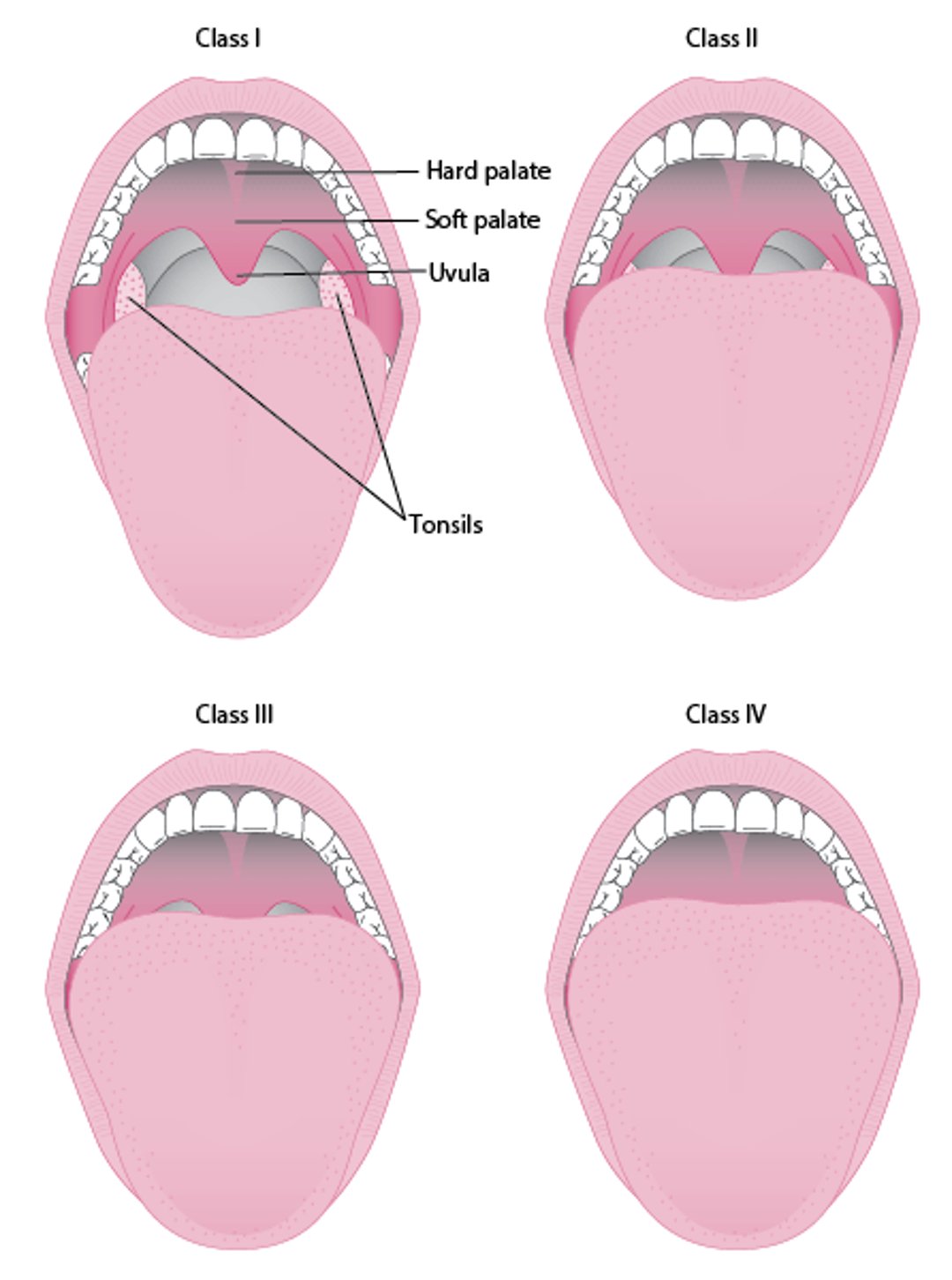
Enlarged tonsils (palatine or lingual), adenoid, tongue, uvula, lateral walls of the pharynx or soft palate (modified Mallampati score 3 or 4—see figure Modified Mallampati Scoring)
Decreased pharyngeal patency
Redundant lateral pharyngeal mucosa
Modified Mallampati Scoring
Modified Mallampati scoring is as follows:
|

The chest should be examined for expiratory wheezes and kyphoscoliosis. Signs of right ventricular failure, including lower-extremity edema, should be noted. A thorough neurologic examination should be performed.
Red flags
The following findings are of particular concern:
Falling asleep while driving or other potentially dangerous situations
Repeated sleep attacks (falling asleep without warning)
Breathing interruptions or awakening with gasping reported by bed partner
Unstable cardiac or pulmonary status
Recent stroke
Status cataplecticus (continuous cataplexy attacks)
History of violent behaviors or injury to self or others during asleep
Frequent sleepwalking or other out-of-bed behavior
Interpretation of findings
Inadequate sleep hygiene and situational stressors are usually apparent in the history. EDS that disappears when sleep time is increased (eg, on weekends or vacations) suggests inadequate sleep syndrome. EDS that is accompanied by cataplexy, hypnagogic/hypnopompic hallucinations, or sleep paralysis suggests narcolepsy.
Difficulty falling asleep (sleep-onset insomnia) should be distinguished from difficulty maintaining sleep and early awakening (sleep maintenance insomnia).
Sleep-onset insomnia suggests delayed sleep phase syndrome, chronic psychophysiologic insomnia, restless legs syndrome, or childhood phobias.
Sleep maintenance insomnia suggests major depression, central sleep apnea, obstructive sleep apnea, periodic limb movement disorder, or aging.
Falling asleep early and awakening early suggest advanced sleep phase syndrome.
Clinicians should suspect obstructive sleep apnea in patients with significant snoring, frequent awakenings, and other risk factors. The STOP-BANG score can help predict risk of obstructive sleep apnea (see table STOP-BANG Risk Score for Obstructive Sleep Apnea).
Testing
Sleep tests are usually performed when specific symptoms or signs suggest obstructive sleep apnea, nocturnal seizures, narcolepsy, periodic limb movement disorder, or other disorders whose diagnosis relies on identification of characteristic polysomnographic findings. Tests are also performed when the clinical diagnosis is in doubt or when response to initial presumptive treatment is inadequate. If symptoms or signs strongly suggest certain causes (eg, restless legs syndrome, poor sleep habits, transient stress, shift work disorder), testing is not required.
Polysomnography is particularly useful when obstructive sleep apnea, narcolepsy, nocturnal seizures, periodic limb movement disorder, or parasomnias are suspected. It also helps clinicians evaluate violent and potentially injurious sleep-related behaviors (REM behavioral disorder). It monitors brain activity (via EEG), eye movements, heart rate, respirations, oxygen saturation, and muscle tone and activity during sleep. Video recording may be used to identify abnormal movements during sleep (1). Polysomnography is typically performed in a sleep laboratory; home sleep studies are now commonly used to diagnose obstructive sleep apnea, but not other sleep disorders (2).
Multiple sleep latency testing assesses speed of sleep onset in 4 to 5 daytime nap opportunities 2 hours apart during the patient’s typical daytime. Patients lie in a darkened room and are asked to sleep. Onset and stage of sleep (including REM) are monitored by polysomnography to determine the degree of sleepiness. This test’s main use is in the diagnosis of narcolepsy.
For the maintenance of wakefulness test, patients are asked to stay awake in a quiet room during 4 wakefulness opportunities 2 hours apart while they sit in a bed or a recliner.
Patients with EDS may require laboratory tests of renal, liver, and thyroid function.
Evaluation references
1. Kushida CA, Littner MR, Morgenthaler T, et al: Practice parameters for polysomnography and procedures: An update for 2005. Sleep 28(4):499-519.
2. Rosen IM, Kirsch DB, Chervin RD, et al: Clinical use of a home sleep apnea test: An American Academy of Sleep Medicine Position Statement. J Clin Sleep Med 13 (10):1205-1207, 2017. doi: 10.5664/jcsm.6774
Treatment of Sleep or Wakefulness Disorders
Specific conditions are treated. The primary treatment for insomnia is cognitive-behavioral therapy (CBT) instead of hypnotics (1), but CBT is not yet widely available for this disorder. Good sleep hygiene, which is important whatever the cause, is a component of cognitive-behavioral therapy and is often the only treatment patients with mild problems need.
Cognitive-behavioral therapy
Cognitive-behavioral therapy for insomnia focuses on managing the common thoughts, worries, and behaviors that interfere with sleep. It is typically performed in 4 to 8 individual or group sessions and can be performed face-to-face, via telehealth remotely, or digitally. Most studies show that telehealth is noninferior to in-person CBT-I (2, 3). However, evidence for the effectiveness of digital CBT-I is weaker (4), although more data are needed to substantiate this.
Cognitive-behavioral therapy for insomnia consists of the following:
Helping patients improve sleep hygiene, particularly restricting time spent in bed, establishing a regular sleep schedule, and controlling stimuli
Teaching patients about the effects of sleeplessness and helping them identify inappropriate expectations about how much sleep they should get
Teaching patients relaxation techniques
Using other cognitive therapy techniques as needed
Restricting the amount of time spent in bed aims to limit the time patients spend lying in bed trying unsuccessfully to sleep. Initially, time in bed is limited to the average nightly total sleep time, but not to < 5.5 hours. Patients are asked to get out of bed in the morning at a fixed time and then calculate a bed time based on total sleep time and remaining awake until that time (called sleep restriction therapy). After a week, this approach typically improves quality of sleep. Then, the time spent in bed can be increased by gradually making bed time earlier, as long as awakenings in the middle of the night remain minimal.
Hypnotics
General guidelines for use of hypnotics (see table Guidelines for the Use of Hypnotics) aim at minimizing abuse, misuse, and addiction.
For commonly used hypnotics, see table Oral Hypnotics in Common Usegamma-aminobutyric (GABA) receptor and augment the inhibitory effects of GABA.
Patients who experience daytime sedation, incoordination, or other daytime effects should avoid activities requiring alertness (eg, driving), and the dose should be reduced, the medication stopped, or, if needed, another medication used. Other adverse effects include amnesia, hallucinations, incoordination, and falls. Falling is a significant risk with all hypnotics.
When benzodiazepines are to be stopped, they should be tapered and not stopped abruptly.
5).
Hypnotics should be used cautiously in patients with pulmonary insufficiency. In older patients, any hypnotic, even in small doses, can cause restlessness, excitement, falls, or exacerbation of delirium and dementia. Rarely, hypnotics can cause complex sleep-related behaviors, such as sleepwalking and even sleep driving; use of higher-than-recommended doses and concurrent consumption of alcoholic beverages may increase risk of such behaviors. Rarely, severe allergic reactions occur.
Prolonged use of hypnoticsWithdrawal and detoxification).
Other medications and substances used to treat insomnia
Many medications and substances not specifically indicated for insomnia are used to induce and maintain sleep.
Some antidepressants
Over-the-counter (OTC) antihistaminesnot be used to treat insomnia. Efficacy is unpredictable, and these agents have long a half-life and have adverse effects such as daytime sedation, confusion, urinary retention, and other systemic anticholinergic effects, which are particularly worrisome in older adults.
Alcohol should also not be used to help with sleep because it produces unrefreshing, disturbed sleep with frequent nocturnal awakenings, often increasing daytime sleepiness. Alcohol can further impair respiration during sleep in patients with obstructive sleep apnea and other pulmonary disorders such as chronic obstructive pulmonary disease (COPD).
Cannabinoids include the following:
CBN (cannabinol), which causes sedation, reduces pain, and increases appetite
THC (tetrahydrocannabinol), which causes euphoria, reduces pain and nausea, and has variable effects on sleep stages
Whether cannabis is effective for insomnia is unclear, but it is useful for chronic pain.
Tolerance can develop; stopping cannabis after long-term use results in insomnia.
Treatment references
1. Qaseem A, Kansagara D, Forciea MA, C et al: Management of Chronic Insomnia Disorder in Adults: A Clinical Practice Guideline From the American College of Physicians. Ann Intern Med 165(2):125-133, 2016. doi: 10.7326/M15-2175
2. Arnedt JT, Conroy DA, Mooney A, et al: Telemedicine versus face-to-face delivery of cognitive behavioral therapy for insomnia: a randomized controlled noninferiority trial. Sleep 44(1):zsaa136, 2021. doi: 10.1093/sleep/zsaa136
3. Gehrman P, Gunter P, Findley J, et al: Randomized noninferiority trial of telehealth delivery of cognitive behavioral treatment of insomnia compared to in-person care. J Clin Psychiatry 82(5):20m13723, 2021. doi: 10.4088/JCP.20m13723
4. Bullock G, Johnson GS, Pattridge SG, et al: A homozygous MAN2B1 missense mutation in a Doberman Pinscher dog with neurodegeneration, cytoplasmic vacuoles, autofluorescent storage granules, and an α-mannosidase deficiency. Genes (Basel)14(9):1746, 2023. doi: 10.3390/genes14091746
5. Zhou M, Tang S: Effect of a dual orexin receptor antagonist on Alzheimer's disease: Sleep disorders and cognition. Front Med (Lausanne) 9:984227, 2023. doi: 10.3389/fmed.2022.984227
6. Cheung JMY, Scott H, Muench A, et al: Comparative short-term safety and efficacy of hypnotics: A quantitative risk-benefit analysis. J Sleep Res e14088, 2023. doi: 10.1111/jsr.14088
Key Points
Poor sleep hygiene and situational disruptors (eg, shift work, emotional stressors) are common causes of insomnia.
Consider medical disorders (eg, sleep apnea syndromes, pain disorders) and psychiatric disorders (eg, mood disorders) as possible causes.
Usually, consider sleep studies (eg, polysomnography) when sleep apnea syndromes, periodic limb movements, or other sleep disorders are suspected, when the clinical diagnosis is in doubt, or when response to initial presumptive treatment is inadequate.
Good sleep hygiene, sometimes as part of cognitive-behavioral therapy, is first-line treatment.
Use hypnotics and sedatives with caution, especially in older adults.


