Growths in the pelvic area (pelvic masses) are common in women.
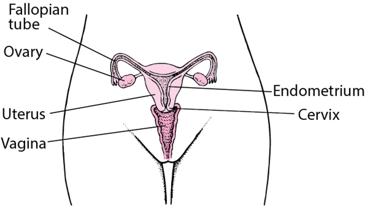
The pelvic area is the lower area of the abdomen. The pelvic area contains the intestines, lower ureters, bladder, and, rarely, a pelvic kidney (kidney in improper position during development of the fetus). The female pelvic area also contains the ovaries, fallopian tubes, uterus, and cervix. A pelvic mass may form in any of these organs.
The types of pelvic masses that may occur vary depending on a woman's age and reproductive phase. Masses during the reproductive years (between the first menstrual period and menopause) in women who are not pregnant and in postmenopausal women are discussed here. Children may develop pelvic masses, which are discussed separately (see Female Pelvic Mass in Children).
A pelvic mass may or may not cause symptoms. Symptoms may include
Dull or sharp pelvic pain that comes and goes (intermittent) or is constant (persistent)
Sudden, severe abdominal pain if a mass bursts (ruptures) or causes the ovary or fallopian tube to twist (a disorder called adnexal torsion)
Menstrual abnormalities
Pain in the lower abdomen during sexual activity
Pelvic masses may be detected during a pelvic examination or with imaging, such as ultrasound or computed tomography (CT).
A pelvic mass is usually noncancerous (benign) but may be cancerous (malignant).
Internal Female Reproductive Anatomy

Evaluation of a Female Pelvic Mass
Doctors ask a woman questions about her symptoms, medical history, and medications. Family history of cancer, especially ovarian, uterine, breast, or colorectal cancer, is important. Doctors also ask about any history of ovarian cysts, uterine fibroids, and history of or risk factors for sexually transmitted infections.
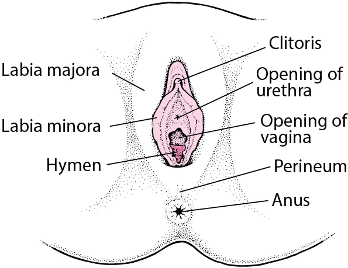
Doctors then do a physical examination, including examination of the abdomen and a pelvic examination. A pelvic examination includes evaluation of the external anatomy (vulva) and internal anatomy (vagina, cervix, uterus, fallopian tubes, ovaries). A combination rectal and vaginal examination may be done. For this examination, the clinician inserts the index finger into the vagina and the middle finger into the rectum. Sometimes masses or abnormalities in the posterior part of the pelvis (the part toward the spine) or in the tissue between the vagina and rectum (perineum) can only be detected with this type of examination.
External Female Reproductive Anatomy

Information from the history and physical examination often suggests a cause and additional tests that may be needed.
A urine or blood pregnancy test is done in women of childbearing age with a pelvic mass. A pregnancy test is done even if pregnancy is unlikely based on menstrual or sexual history. An undiagnosed pregnancy increases the risk of complications to the woman or fetus. If a pregnancy test is positive, the most likely cause of the mass is the enlarged pregnant uterus.
Imaging, such as a pelvic ultrasound, is often done if doctors suspect a pelvic mass. (See also Gynecologic Imaging Studies.)
If a cervical mass is found during the pelvic examination, additional tests may include a Papanicolaou (Pap) test and biopsy.
Postmenopausal women may require additional evaluation, because the risk of a pelvic mass being cancerous increases with age.
Treatment of a Female Pelvic Mass
After a pelvic mass is detected, clinicians determine if a woman requires urgent treatment (for example, for ectopic pregnancy, ovarian torsion, or a ruptured ovarian cyst with severe bleeding). Doctors also determine if the mass may be cancerous. Severe pain often requires urgent treatment.
Some masses are monitored and may not need treatment or may resolve without treatment. For some noncancerous masses, symptoms can be managed with medications or minimally invasive procedures (such as uterine fibroid embolization). However, in some cases, pelvic masses require surgical treatment or removal.
Key Points
A female pelvic mass may originate from the ovaries, fallopian tubes, uterus, or cervix, as well as the intestines, lower ureters, bladder, or, rarely, a pelvic kidney.
In women of reproductive age, common causes of pelvic masses are uterine fibroids and follicular ovarian cysts.
In postmenopausal women, masses are more likely to be cancerous than in younger women.
A pelvic mass may or may not cause symptoms and may be detected during pelvic examination or with an imaging test. A pelvic mass is usually noncancerous (benign) but may be cancerous (malignant).
Women of childbearing age with a pelvic mass receive a pregnancy test, even if pregnancy is unlikely.
Doctors use imaging tests to evaluate women with a pelvic mass.