


(See also Overview of Plasma Cell Disorders.)
In the United States, the lifetime risk of getting multiple myeloma is 1 in 132 (0.76%) (1). The median age is about 70 years. Prevalence in Black people is twice that in White people. There is a slight male predominance. The etiology is unknown, although chromosomal and genetic factors, radiation, and chemicals have been suggested.
The American Cancer Society estimates that in 2024 in the United States there will be about 35,780 new cases of multiple myeloma diagnosed and about 12,540 deaths (1). Survival continues to improve with recent reports showing a median survival of over 10 years (2).
General references
1. American Cancer Society. Key Statistics About Multiple Myeloma. Atlanta, Ga., American Cancer Society; 2024.
2. Jew S, Bujarski S, Rgidor B, et al: Clinical outcomes and serum B-cell maturation antigen levels in a real-world unselected population of newly diagnosed multiple myeloma patients. Target Oncol 18(5):735–747, 2023. doi:10.1007/s11523-023-00990-6
Pathophysiology of Multiple Myeloma
The M-protein (monoclonal immunoglobulin protein) produced by the malignant plasma cells is IgG in about 50 to 55% of myeloma patients and IgA in about 20% (1). Of patients producing either IgG or IgA, 40% also have Bence Jones proteinuria, which is free monoclonal kappa (κ) or lambda (λ) light chains in the urine. In 15 to 20% of patients, plasma cells secrete only Bence Jones protein. IgM and IgD myeloma account for approximately 1 to 2% of cases each; IgD myeloma is more frequent among patients of Asian descent. IgE myeloma is exceedingly rare.
Rarely, patients have no M-protein in blood and urine. However, the serum free light chain assay demonstrates monoclonal light chains in many of the patients who were formerly considered to have nonsecretory myeloma.
Diffuse osteoporosis or discrete osteolytic lesions are often present, with the most common sites being the pelvis, spine, ribs, femur, humerus, and skull. Lesions are caused by bone replacement by expanding plasmacytomas or by cytokines that are secreted by malignant plasma cells that activate osteoclasts and suppress osteoblasts, leading to bone loss. The osteolytic lesions are usually multiple; occasionally, they are solitary intramedullary masses. Increased bone loss may also lead to hypercalcemia.
Extraosseous solitary plasmacytomas are unusual but may occur in any tissue, especially in the upper respiratory tract.
In many patients, renal failure is present at diagnosis or develops during the course of the disorder. Renal failure has many causes, and most commonly, it results from deposition of light chains in the distal tubules (myeloma-related kidney disease) or hypercalcemia.
Patients often develop anemia usually due to kidney disease or suppression of erythropoiesis by cancer cells. Anemia in patients with multiple myeloma also may be due to other unrelated causes, including iron deficiency or vitamin B12 deficiency.
Because of lack of normal antibodies and other immune dysfunction, some patients have increased susceptibility to bacterial infection. Viral infections, especially herpes zoster infections
Amyloidosis occurs in 10% of patients who have myeloma, most often in patients with lambda-type M-proteins (2).
Variant expressions of multiple myeloma occur (see table Variant Expressions of Multiple Myeloma).
Variant Expressions of Multiple Myeloma
Form | Characteristics |
|---|---|
Extramedullary plasmacytoma | Plasmacytomas that occur outside of the bone marrow |
Solitary plasmacytoma of bone | Single bone plasmacytomas, which usually produce no M-protein |
Osteosclerotic myeloma (POEMS syndrome) | Polyneuropathy (chronic inflammatory polyneuropathy) Organomegaly (hepatomegaly, splenomegaly, or lymphadenopathy) Endocrinopathy (eg, gynecomastia, testicular atrophy) M-protein Skin changes (eg, hyperpigmentation, excess hair) |
Nonsecretory myeloma | Absence of M-protein in serum and urine Absence of elevated serum free light chains |
Pathophysiology references
1. Kyle RA, Gertz MA, Witzig TE, et al. Review of 1027 patients with newly diagnosed multiple myeloma. Mayo Clin Proc 2003;78(1):21-33. doi:10.4065/78.1.21
2. Ríos-Tamayo R, Krsnik I, Gómez-Bueno M, et al. AL Amyloidosis and Multiple Myeloma: A Complex Scenario in Which Cardiac Involvement Remains the Key Prognostic Factor. Life (Basel). 2023;13(7):1518. Published 2023 Jul 6. doi:10.3390/life13071518
Symptoms and Signs of Multiple Myeloma
Persistent bone pain (especially in the back or thorax), renal failure, and recurrent bacterial infections are the most common problems on presentation, but many patients are identified when routine laboratory tests show an elevated total protein level in the blood, proteinuria, or unexplained anemia or renal failure.
Symptoms of anemia predominate or may be the sole reason for evaluation in some patients, and a few patients have manifestations of the hyperviscosity syndrome.
Pathologic (fragility) fractures (ie, fractures that occur with minimal or no trauma) are common, and vertebral collapse may lead to spinal cord compression and paraplegia.
Peripheral neuropathy, carpal tunnel syndrome (especially in patients with amyloidosis), abnormal bleeding, and symptoms of hypercalcemia (eg, polydipsia, dehydration) are common.
Lymphadenopathy and hepatosplenomegaly are unusual.
Diagnosis of Multiple Myeloma
Complete blood count (CBC) with platelets, peripheral blood smear, and chemistry panel (blood urea nitrogen [BUN], creatinine, calcium, uric acid, lactate dehydrogenase [LDH])
Serum and urine protein (from a 24-hour urine collection) electrophoresis followed by immunofixation; quantitative immunoglobulins; and serum free light chains
Radiographs (skeletal survey) and either a positron emission tomography (PET)-CT scan or whole-body MRI
Bone marrow examination, including conventional cytogenetics and fluorescent in situ hybridization studies (FISH)
Multiple myeloma is suspected in patients > 40 years with persistent unexplained bone pain, particularly at night or at rest, other typical symptoms, unexplained laboratory abnormalities (eg, elevated blood protein or urinary protein, hypercalcemia, renal insufficiency, anemia), or radiographs showing a pathologic fracture or lytic lesions.
Laboratory evaluation includes routine blood tests, LDH, serum beta-2 microglobulin, urine and serum immunofixation and protein electrophoresis, and serum free light chains. Patients should also undergo a skeletal survey and either a PET-CT scan or whole-body MRI because these tests are more sensitive to bone disease than radiographs. A bone marrow examination is also required along with conventional cytogenetics and FISH studies (for review, see references 1 and 2).
Routine blood tests include CBC and chemistry panel. Anemia is present in 80% of patients, usually a normocytic, normochromic anemia with formation of rouleaux (clusters of 3 to 12 red blood cells that occur in stacks) (3). White blood cell and platelet counts are usually normal. BUN, serum creatinine, LDH, beta-2 microglobulin, and serum uric acid may be elevated. The anion gap is sometimes low. Hypercalcemia is present at diagnosis in about 10% of patients.
Protein and immunofixation electrophoresis are carried out on a serum sample and on a urine sample concentrated from a 24-hour collection to identify, quantify, and characterize M-protein. Serum protein electrophoresis identifies M-protein in about 80 to 90% of patients. Most of the remaining 10 to 20% are patients with only free monoclonal light chains (Bence Jones protein) that can be detected in blood using a free light chain assay or in urine using urine protein and immunofixation electrophoresis. Monoclonal protein is not evident at diagnosis in a small proportion of patients; during the course of the disease, fewer patients have evidence of monoclonal protein.
Immunofixation electrophoresis can identify the immunoglobulin class of the M-protein (IgG, IgA, or uncommonly IgD, IgM, or IgE) and can often detect light-chain protein if serum immunoelectrophoresis is negative; immunofixation electrophoresis is done even when the serum test is negative if multiple myeloma is strongly suspected.
Serum free light-chain analysis with delineation of kappa and lambda ratios or differences between the involved and uninvolved light chains helps confirm the diagnosis and can also be used to monitor efficacy of therapy and provide prognostic data.
Serum level of beta-2 microglobulin is measured if the diagnosis is confirmed or very likely, and along with serum albumin, is used as part of the international staging system (see table Revised International Staging System for Multiple Myeloma). Beta-2 microglobulin is a small protein on the membrane of all cells. Its concentration varies directly with tumor mass and severity of renal dysfunction.
Revised International Staging System for Multiple Myeloma
Stage | Criteria |
|---|---|
I | Beta-2 microglobulin < 3.5 mcg/mL (< 297 nmol/L) and Serum albumin ≥ 3.5 g/dL (≥ 35 g/L) Normal LDH Standard-risk cytogenetic abnormalities by FISH* |
II | Not stage I or III |
III | Beta-2 microglobulin ≥ 5.5 mcg/mL (≥ 466 nmol/L) and High-risk cytogenetic abnormalities by FISH† and/or high LDH |
* Standard-risk cytogenetic abnormalities include t(11;14), t(6;14), and trisomies. | |
† High-risk cytogenetic abnormalities consist of t(4;14), t(14;16), or 17p-. | |
FISH = fluorescent in situ hybridization; LDH = lactate dehydrogenase. | |
Data from Palumbo A, Avet-Loiseau H, Oliva S, et al. Revised International Staging System for Multiple Myeloma: A Report From International Myeloma Working Group. J Clin Oncol 2015;33(26):2863-2869. doi:10.1200/JCO.2015.61.2267 | |
Radiographs include a skeletal survey (ie, plain radiographs of skull, long bones, spine, pelvis, and ribs). Punched-out lytic lesions or diffuse osteoporosis is present in approximately 80% of cases (3). Radionuclide bone scans usually are not helpful. Whole-body MRI can provide more detail and is obtained if specific sites of pain or neurologic symptoms are present. PET-CT scans provide prognostic information and can help determine whether patients have solitary plasmacytoma or multiple myeloma.

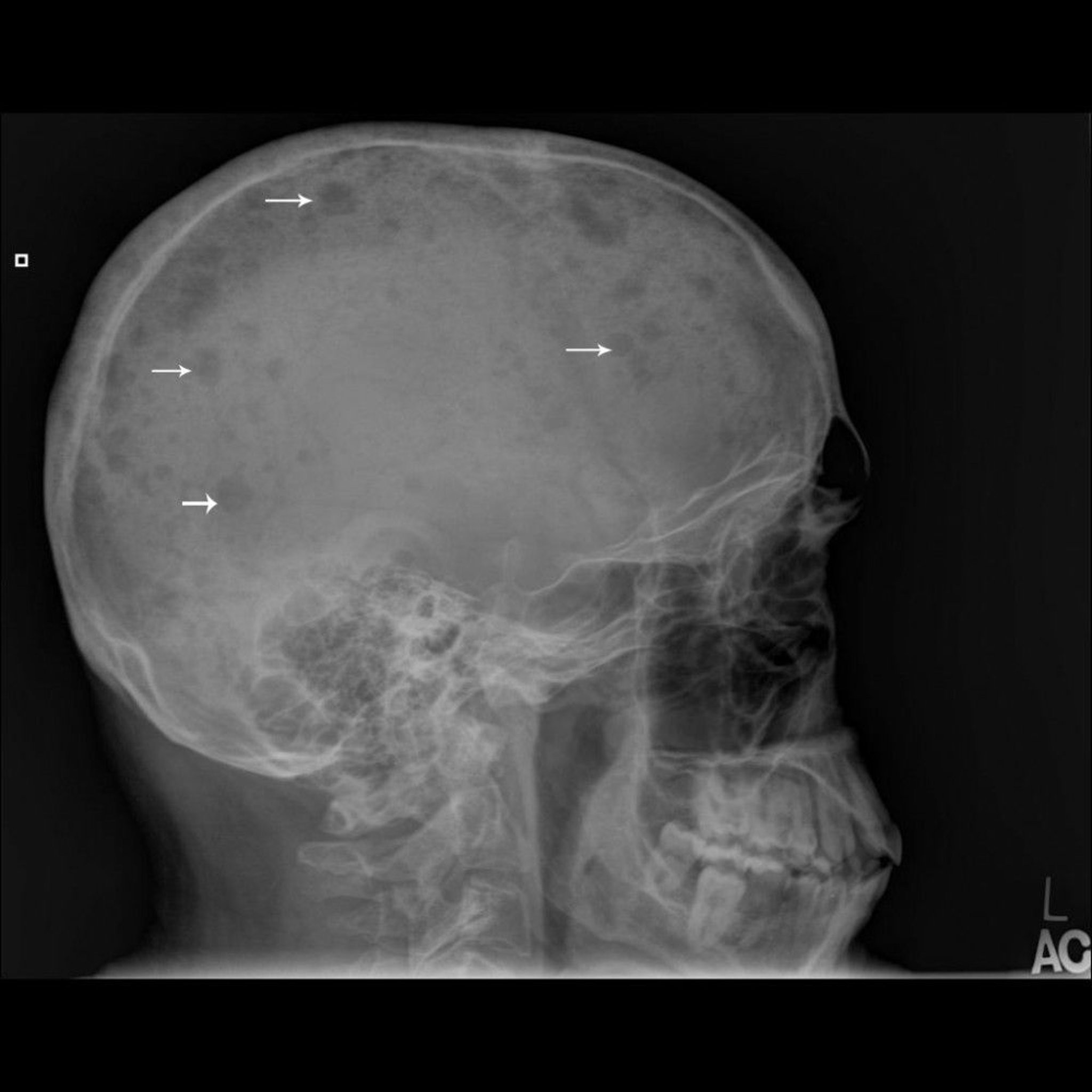
Image courtesy of Michael J. Joyce, MD, and Hakan Ilaslan, MD.
Bone marrow aspiration and biopsy are done and reveal sheets or clusters of plasma cells; myeloma is diagnosed when ≥ 10% of the cells are of this type. However, bone marrow involvement is patchy; therefore, some samples from patients with myeloma may show < 10% plasma cells. Still, the percentage of plasma cells in bone marrow is rarely normal. Plasma cell morphology does not correlate with the class of immunoglobulin synthesized. Chromosomal studies on bone marrow (eg, using cytogenetic testing methods such as FISH and immunohistochemistry) may reveal specific karyotypic abnormalities in plasma cells that can influence treatment choices and are associated with differences in survival.
Diagnosis and differentiation from other malignancies (eg, metastatic carcinoma, lymphoma, leukemia) and from monoclonal gammopathy of undetermined significance typically require multiple criteria:
Clonal bone marrow plasma cells or plasmacytoma
M-protein in plasma and/or urine
Organ impairment (hypercalcemia, renal insufficiency, anemia, or bony lesions)
In patients without serum M protein, myeloma is indicated by Bence Jones proteinuria > 200 mg/24 hour or abnormal serum free light chain levels, osteolytic lesions (without evidence of metastatic cancer or granulomatous disease), and sheets or clusters of plasma cells in the bone marrow.
Diagnosis references
1. Sive J, Cuthill K, Hunter H, Kazmi M, Pratt G, Smith D and on behalf of British Society of Haematology: Guidelines on the diagnosis, investigation and initial treatment of myeloma: a British Society for Haematology/UK Myeloma Forum Guideline. Brit J Haematol 193:245–268, 2021. doi:10.1111/bjh.17410
2. Rajkumar SV: Multiple myeloma: 2022 update on diagnosis, risk-stratification and management. Am J Hematol 97(8):1086-1107, 2022. doi:10.1002/ajh.26590
3. Kyle RA, Gertz MA, Witzig TE, et al. Review of 1027 patients with newly diagnosed multiple myeloma. Mayo Clin Proc. 2003;78(1):21-33. doi:10.4065/78.1.21
Treatment of Multiple Myeloma
Conventional chemotherapy and corticosteroids for patients with symptoms
Additional therapy with immunomodulatory agents and monoclonal antibodies
Possibly autologous stem cell transplantation
Possibly radiation therapy to specific symptomatic areas that do not respond to systemic therapy
Treatment of complications, eg, anemia, hypercalcemia, kidney dysfunction, infections, and bone disease
Treatment of relapsed or refractory disease with immunomodulatory agents, monoclonal antibodies, proteasome inhibitors, and newer cellular treatments
Therapy involves direct treatment of malignant cells in patients with symptoms and those with myeloma-related organ dysfunction (anemia, renal dysfunction, hypercalcemia, or bone disease).
Among patients initially presenting without organ dysfunction, risk factors for requiring rapid treatment of myeloma include
> 60% plasma cells in bone marrow
> 1 lesion on MRI
Serum free light chain levels > 100 mg/L
Patients with these risk factors are considered to have active myeloma and require immediate treatment even though early treatment of these patients has not been shown to improve their overall survival. Patients without these risk factors or end-organ dysfunction probably do not benefit from immediate treatment, which is usually withheld until symptoms or complications develop.
Treatment of malignant cells
Conventional chemotherapy
1).
Response to treatment (see table Defining Response to Cancer Treatment) is indicated by
Decreases in serum and urine M-protein
Decreases in levels of the involved serum free light chain
Increases in numbers of red blood cells
Improvement in renal function among patients presenting with renal failure
Normalization of calcium levels among those presenting with elevated levels
Decrease in bone pain
Decrease in fatigue
Autologous peripheral blood stem cell transplantation may be considered for patients who have adequate cardiac, hepatic, pulmonary, and renal function, particularly those whose disease is stable or responsive after several cycles of initial therapy. However, pharmacotherapy options are highly effective and may make transplantation less often necessary or unnecessary altogether. Clinical trials show longer progression-free survival, but no improvement in overall survival when patients undergo stem cell transplantation as part of initial therapy (2).
Treatment of relapsed or refractory myeloma
For patients with relapsed or refractory myeloma, effective combinations include
Corticosteroids
These medications are usually combined with other effective agents that the patient has not yet been treated with.
Patients with prolonged remissions may respond to retreatment with the same regimen that led to the initial remission. Patients who fail to respond to a given combination of medications may respond when another medication in the same class (eg, proteasome inhibitors, immunomodulatory agents, chemotherapeutic agent) is substituted.
Monoclonal antibodies targeting proteins on myeloma cells are also highly effective in relapsed or refractory myeloma. Available monoclonal antibodies include those that target
Effective immune treatments that target B-cell maturation antigen (BCMA) are available. These treatments include
Although these treatments are effective, they can cause significant acute adverse effects (cytokine release syndrome, neurologic problems), a high risk of ongoing severe infections, and secondary cancers.
7).
Maintenance therapy
Maintenance therapy has been tried with nonchemotherapeutic medications, including interferon alfa, which prolongs remission but does not improve survival and is associated with significant adverse effects. Following a response to corticosteroid-based regimens, corticosteroids alone are effective as a maintenance treatment.
autologous stem cell transplantation. Thus, the risk of developing secondary cancers must be weighed against improved survival.
The role of monoclonal antibodies as maintenance therapy remains to be defined.
Treatment of complications
In addition to direct treatment of malignant cells, therapy must also be directed at complications, which include
Anemia
Hypercalcemia
Hyperuricemia
Hyperviscosity
Infections
Renal insufficiency
Skeletal lesions
Anemia that is inadequately relieved by chemotherapy can be treated with recombinant erythropoietin. If anemia causes cardiovascular symptoms or significant systemic symptoms, packed red blood cells are transfused. Often patients are iron deficient for reasons unrelated to their myeloma and require intravenous iron. Patients with anemia should have periodic measurement of serum iron, transferrin, and ferritin levels to monitor iron stores as well as vitamin B12 levels.
If hyperviscosity develops, which rarely occurs in patients with myeloma, plasma exchange is indicated.
Hypercalcemiavitamin D.
Hyperuricemiatumor lysis syndrome with treatment.
Infection is more likely during treatment-induced neutropenia. In addition, infections with the herpes zoster
Documented bacterial infections should be treated with antibiotics. Prophylactic use of antibiotics is not routinely recommended.
Prophylactic IV immune globulin may reduce the risk of infection but is generally reserved for patients with low uninvolved immunoglobulin levels and frequent recurrent infections.
and influenza vaccine are indicated to prevent infection but are not effective in most patients because of disease-related and treatment-related immune deficiency. Use of live vaccines is not recommended in patients with a compromised immune system. To prevent herpes zoster infection, the nonviable recombinant zoster vaccine, may be given but is of limited effectiveness.
Renal compromise can often be ameliorated with adequate hydration. Even patients with prolonged, massive Bence Jones proteinuria (≥ 10 to 30 g/day) may have intact renal function if they maintain a urine output > 2000 mL/day. Dehydration combined with high-osmolar IV contrast may precipitate acute oliguric renal failure in patients with Bence Jones proteinuria. Plasma exchange may be effective in some cases. Nephrotoxic medications should be avoided. Rapid and aggressive treatment of the underlying myeloma to reduce the levels of the nephrotoxic monoclonal immunoglobulin is important to reverse this condition.
Skeletal lesions require multiple supportive measures. Maintenance of ambulation and supplemental calcium and vitamin D, as indicated, help preserve bone density in patients without disease-related hypercalcemia. Vitamin D levels should be measured at diagnosis and periodically, and dosing of vitamin D adjusted accordingly. Analgesics and palliative doses of radiation therapy (18 to 24 gray) can relieve bone pain. However, radiation therapy may cause significant toxicity and, because it suppresses bone marrow function, may impair the patient’s ability to receive cytotoxic doses of systemic chemotherapy.
Treatment references
1. Abdallah N, Kumar SK. Daratumumab in untreated newly diagnosed multiple myeloma. Ther Adv Hematol 2019;10:2040620719894871. doi:10.1177/2040620719894871
2. Richardson PG, Jacobus SJ, Weller EA, et al. Triplet Therapy, Transplantation, and Maintenance until Progression in Myeloma. N Engl J Med 2022;387(2):132-147. doi:10.1056/NEJMoa2204925
3. Palumbo A, Chanan-Khan A, Weisel K, et al. Daratumumab, Bortezomib, and Dexamethasone for Multiple Myeloma. N Engl J Med 2016;375(8):754-766. doi:10.1056/NEJMoa1606038
4. Arnall JR, Maples KT, Harvey RD, et al. Daratumumab for the treatment of multiple myeloma: A review of clinical applicability and operational considerations. AnnalsPharmacother 2022;56(8):927-940. doi: 10.1177/10600280211058754
5. Yang J, Zhou W, Li D, et al. BCMA-targeting chimeric antigen receptor T-cell therapy for multiple myeloma. Cancer Lett 2023;553:215949. doi: 10.1016/j.canlet.2022.215949
6. Lee H, Neri P, Bahlis NJ. BCMA- or GPRC5D-targeting bispecific antibodies in multiple myeloma: efficacy, safety, and resistance mechanisms. Blood 2024;143(13):1211-1217. doi: 10.1182/blood.2023022499
7. He W, He F, Hu H. Efficacy and safety of venetoclax-based regimens in relapsed or refractory multiple myeloma: a systematic review and meta-analysis of prospective trials. Ann Med 2023;55(1):1029-1036. doi:10.1080/07853890.2023.2186480
Prognosis for Multiple Myeloma
Although multiple myeloma is progressive and remains incurable, the median overall survival has improved to more than 11 years among unselected patients in a recent study (1).
Because multiple myeloma is ultimately fatal, patients are likely to benefit from discussions of end-of-life care that involve clinicians and appropriate family and friends. Points for discussion may include advance directives, the use of feeding tubes, and pain relief.
Prognosis reference
1. Jew S, Bujarski S, Regidor B, et al. Clinical outcomes and serum B-cell maturation antigen levels in a real-world unselected population of newly diagnosed multiple myeloma patients. Target Oncol 2023;18:735-747. doi: 10.1007/s11523-023-00990-6
Key Points
Malignant plasma cells produce monoclonal immunoglobulin and invade and destroy bone.
Expanding plasmacytomas and cytokine secretion cause multiple, discrete, osteolytic lesions (usually in the pelvis, spine, ribs, and skull) and diffuse osteoporosis; pain, fractures, and hypercalcemia are common.
Anemia and renal failure are common.
Amyloidosis develops in about 10%, typically patients who produce excess lambda light chains.
Do serum and urine protein electrophoresis followed by immunofixation, quantitative immunoglobulins, and measurement of serum free light chains.
Do bone marrow aspiration and biopsy.
Patients with symptoms and those with organ dysfunction should be treated with pharmacotherapy, which may include corticosteroids, chemotherapy agents, proteasome inhibitors, immunomodulatory agents, monoclonal antibodies, selective inhibitors of nuclear export, histone deacetylase inhibitors, and cellular and antibody-based immune therapies targeting B-cell maturation antigen.
Stem cell transplantation is an option for some patients, but highly effective pharmacotherapy options may make it unnecessary in others.
More Information
The following English-language resources may be useful. Please note that THE MANUAL is not responsible for the content of these resources.
Bal S, Giri S, Godby KN, Costa LJ: New regimens and directions in the management of newly diagnosed multiple myeloma. Am J Hematol 96:367–378, 2021. doi:10.1002/ajh.26080
Cook G, Morris CTCM: Evolution or revolution in multiple myeloma therapy and the role of the UK. Brit J Haematol 191:542–551, 2020. doi:10.1111/bjh.17148
Cowan AJ, Green DJ, Kwok M, et al. Diagnosis and management of multiple myeloma: A review. JAMA 327:464-477, 2022. doi: 10.1001/jama.2022.0003
Gulla A, Anderson KC: Multiple myeloma: the (r)evolution of current therapy and a glance into the future. Haematologica 105:2358–2367, 2020. doi:10.3324/haematol.2020.247015
Rafae A., Rhee FV, Hadidi SA: Perspectives on the treatment of multiple myeloma. Oncologist 29:200-212, 2024. doi: 10.1093/oyad306
Rajkumar SV: Multiple Myeloma: 2022 update on diagnosis, risk stratification, and management. Am J Hematol 97:1086–1107, 2022. doi: 10.1002/ajh.26590


