


Stasis dermatitis is inflammation, typically of the skin of the lower legs, caused by chronic edema. Symptoms are itching, scaling, and hyperpigmentation. Ulceration can be a complication. Diagnosis is clinical. Treatment is directed at the causes of edema and preventing ulceration.
")
(See also Definition of Dermatitis.)
Stasis dermatitis occurs in patients with chronic edema due to a number of conditions, including chronic venous insufficiency, right heart failure, or lymphedema. Increased capillary pressure with subsequent compromise of endothelial integrity in the microvasculature results in fibrin leakage, and disruption of the epithelial barrier function results in local inflammation. Stasis dermatitis occurs most commonly on the shins but can also affect other areas with chronic edema, such as the arms after radiation treatment of axillary lymph nodes.
Stasis dermatitis as well as leg ulcers, which commonly accompany stasis dermatitis in patients with chronic venous insufficiency, are sometimes treated with various topical medications. Thus, contact dermatitis often complicates stasis dermatitis (1).
General reference
1. Erfurt-Berge C, Geier J, Mahler V. The current spectrum of contact sensitization in patients with chronic leg ulcers or stasis dermatitis: New data from the Information Network of Departments of Dermatology (IVDK). Contact Dermatitis. 77(3):151–158, 2017. doi: 10.1111/cod.12763
Symptoms and Signs of Stasis Dermatitis
Manifestations of stasis dermatitis include pruritus, ill-defined erythema, scaling, and lichenification, most commonly on the shins. Plaques may also occur, often weeping and crusted, and can be associated with bacterial superinfection.
When chronic venous insufficiency is the cause, other manifestations usually include varicose veins, purpura jaune d'ocre (a yellow-brown discoloration due to hemosiderin deposits in the dermis), and lipodermatosclerosis (sclerosis of subcutaneous fat caused by panniculitis, also called sclerosing panniculitis), giving the lower leg an inverted bowling pin shape with enlargement of the calf and narrowing at the ankle.

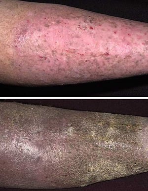
Chronic stasis dermatitis may appear as fibrotic skin thickening and hyperpigmentation. The changes are characteristic in both light skin (top) and dark skin (bottom), here appearing more pronounced in the bottom photo.
Images provided by Thomas Habif, MD.


This photo shows a central large weeping erosion at high risk of developing into a chronic leg ulcer. It is surrounded by chronic changes of venous insufficiency with hyperpigmentation and thickened fibrotic skin.
Roberto A. Penne-Casanova/SCIENCE PHOTO LIBRARY

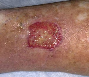
Venous stasis ulcers develop as a result of inadequately treated stasis dermatitis; they may quickly follow the first signs of stasis dermatitis.
Image provided by Thomas Habif, MD.
Chronic stasis dermatitis may appear as fibrotic skin thickening and hyperpigmentation. The changes are characteristic in both light skin (top) and dark skin (bottom), here appearing more pronounced in the bottom photo.
Images provided by Thomas Habif, MD.
This photo shows a central large weeping erosion at high risk of developing into a chronic leg ulcer. It is surrounded by chronic changes of venous insufficiency with hyperpigmentation and thickened fibrotic skin.
Roberto A. Penne-Casanova/SCIENCE PHOTO LIBRARY
Venous stasis ulcers develop as a result of inadequately treated stasis dermatitis; they may quickly follow the first signs of stasis dermatitis.
Image provided by Thomas Habif, MD.
Diagnosis of Stasis Dermatitis
Clinical evaluation
Diagnosis of stasis dermatitis is clinical based on the characteristic appearance of the skin lesions and other signs of chronic leg swelling and venous insufficiency.
Consultation with a vascular specialist and testing (such as Doppler ultrasound) may be needed.
Treatment of Stasis Dermatitis
Treatment of the causes of swelling
Compression and elevation
Treatment of complications (eg, secondary infection, allergic contact dermatitis, ulcers)
The cause of the chronic swelling should be corrected to the extent possible. Leg elevation and compression are often indicated. Chronic venous insufficiency should be treated.
In addition, noneroded stasis dermatitis often abates with a midpotency topical corticosteroid (eg, triamcinolone acetonide 0.1% cream or ointment). For In addition, noneroded stasis dermatitis often abates with a midpotency topical corticosteroid (eg, triamcinolone acetonide 0.1% cream or ointment). Foran eroded (weeping) lesion, a hydrocolloid dressing may be best.
Ulcers are best treated with compresses and bland dressings (eg, zinc oxide paste); other dressings (eg, hydrocolloids) are also effective (see also are best treated with compresses and bland dressings (eg, zinc oxide paste); other dressings (eg, hydrocolloids) are also effective (see alsoDirect wound care).
Oral antibiotics (eg, cephalosporins, dicloxacillin) are used to treat superimposed Oral antibiotics (eg, cephalosporins, dicloxacillin) are used to treat superimposedcellulitis. Topical antibiotics (eg, mupirocin, silver sulfadiazine) are useful for treating superinfected erosions and ulcers. . Topical antibiotics (eg, mupirocin, silver sulfadiazine) are useful for treating superinfected erosions and ulcers.
Complex or multiple topical medications or over-the-counter remedies should not be used. The skin in stasis dermatitis is more vulnerable to direct irritants and to potentially sensitizing topical agents (eg, antibiotics; anesthetics; vehicles of topical medications, especially lanolin or wool alcohols).Complex or multiple topical medications or over-the-counter remedies should not be used. The skin in stasis dermatitis is more vulnerable to direct irritants and to potentially sensitizing topical agents (eg, antibiotics; anesthetics; vehicles of topical medications, especially lanolin or wool alcohols).
Key Points
Stasis dermatitis, most typically on the shins, results from chronic edema.
Signs include erythema, scaling, pruritus, and lichenification and may include weeping erosions and crusting.
Complications include secondary infections, ulcers, and contact sensitivities.
Elevation and compression are often required.


