


Aortic stenosis (AS) is narrowing or restricted opening of the aortic valve, obstructing blood flow from the left ventricle to the aorta during systole. Causes include a congenital bicuspid valve, idiopathic degenerative sclerosis with calcification, and rheumatic fever. Untreated AS progresses to become symptomatic with one or more of the classic triad of syncope, angina, and exertional dyspnea; heart failure and arrhythmias may develop. A crescendo-decrescendo ejection murmur is characteristic. Diagnosis is by physical examination and echocardiography. Asymptomatic AS in adults usually requires no treatment. Once symptoms develop, surgical or percutaneous valve replacement is required. For severe or symptomatic AS in children, balloon valvuloplasty may be effective, but surgery is often required.
(See also Overview of Cardiac Valvular Disorders.)
Etiology of Aortic Stenosis
In older patients, the most common precursor to aortic stenosis is:
Aortic sclerosis
Aortic sclerosis is the deposition of calcium on the aortic valve leaflets, seen in 25 to 45% of adults over age 65 (1, 2). The cause of aortic sclerosis and stenosis is not yet known but is mediated by an inflammatory process similar to but distinct from atherosclerosis. Genetic, anatomic, fluid dynamic, and environmental risk factors include hypertension, smoking, high cholesterol, and presence of a bicuspid valve. Lipid deposition and inflammation initially lead to thickening of aortic valve structures by fibrosis and calcification without causing significant obstruction. Aortic sclerosis progresses to severe aortic stenosis in approximately 10% of patients within 5 years (3).
Lipoprotein (a) is implicated in the pathogenesis of both aortic stenosis and atherosclerosis. Elevated lipoprotein (a) levels also predict faster hemodynamic progression of AS.
Patients with psoriasis are also at increased risk for atherosclerosis, and more recently, psoriasis has been tied to an increased risk of aortic stenosis.
In patients < 70 years, the most common cause of aortic stenosis is:
A congenital bicuspid aortic valve
Congenital aortic valve disease, including bicuspid aortic valve and aortic stenosis, occurs in 0.5 to 2% of people and affects more males (4); it is associated with coarctation and progressive dilatation of the ascending aorta, causing aortic dissection. Aortic stenosis in neonates can also accompany hypoplastic left heart syndrome and its variants.
In medically underserved countries, the most common cause of aortic stenosis in all age groups is:
Supravalvular AS caused by a discrete congenital membrane or narrowing of the sinotubular junction or ascending aorta is uncommon. Supravalvular AS can be a component of Williams syndrome, a microdeletion syndrome of chromosome 7 associated with a characteristic facial appearance (high and broad forehead, hypertelorism, strabismus, upturned nose, long philtrum, wide mouth, dental abnormalities, puffy cheeks, micrognathia, low-set ears) and neonatal hypercalcemia.
Subvalvular AS caused by a congenital membrane or fibrous ring just beneath the aortic valve is uncommon. Hypertrophic cardiomyopathy can have dynamic obstruction of the left ventricular outflow tract.
Etiology references
1. Ferreira-González I, Pinar-Sopena J, Ribera A, et al. Prevalence of calcific aortic valve disease in the elderly and associated risk factors: a population-based study in a Mediterranean area. Eur J Prev Cardiol 2013;20(6):1022-1030. doi:10.1177/2047487312451238
2. Stewart BF, Siscovick D, Lind BK, et al. Clinical factors associated with calcific aortic valve disease. Cardiovascular Health Study. J Am Coll Cardiol 1997;29(3):630-634. doi:10.1016/s0735-1097(96)00563-3
3. Otto CM, Nishimura RA, Bonow RO, et al: 2020 ACC/AHA Guideline for the Management of Patients With Valvular Heart Disease: Executive Summary: A Report of the American College of Cardiology/American Heart Association Joint Committee on Clinical Practice Guidelines. Circulation 143(5):e35–e71, 2021. doi: 10.1161/CIR.0000000000000932
4. Siu SC, Silversides CK. Bicuspid aortic valve disease. J Am Coll Cardiol 2010;55(25):2789-2800. doi:10.1016/j.jacc.2009.12.068
Pathophysiology of Aortic Stenosis
Aortic stenosis causes an increased pressure load on the left ventricle (LV), resulting in compensatory hypertrophy without cavity enlargement (concentric hypertrophy). In early disease stages, there is an increased pressure gradient from the high-pressure LV to the lower pressure aorta. With time, the ventricle can no longer compensate and begins to fail, with secondary LV cavity enlargement, reduced ejection fraction (EF), decreased cardiac output, and a misleadingly low gradient across the aortic valve (low-gradient severe AS) as the LV can no longer generate as high of a systolic pressure.
Aortic regurgitation accompanies aortic stenosis in approximately 7% of patients (1), and about half of patients with severe AS also have mitral annular calcification (2, 3), which may lead to mitral regurgitation.
Patients with other disorders that also cause LV enlargement and reduced EF (eg, myocardial infarction, intrinsic cardiomyopathy) may generate insufficient flow to fully open a sclerotic valve and have an apparently small valve area even when their AS is not particularly severe (pseudo-severe AS). Pseudo-severe AS must be differentiated from low-gradient severe AS because only patients with low-gradient severe AS benefit from valve replacement.


MONICA SCHROEDER / SCIENCE PHOTO LIBRARY
Elevated shear stress across the stenosed aortic valve degrades von Willebrand factor multimers. The resulting coagulopathy may cause gastrointestinal bleeding in patients with angiodysplasia (Heyde syndrome). The syndrome typically resolves following successful aortic valve replacement (4).
Pathophysiology references
1. Nedadur R, Belzile D, Farrell A, Tsang W. Mixed aortic stenosis and regurgitation: a clinical conundrum. Heart 2023;109(4):264-275. Published 2023 Jan 27. doi:10.1136/heartjnl-2021-320501
2. Abramowitz Y, Kazuno Y, Chakravarty T, et al. Concomitant mitral annular calcification and severe aortic stenosis: prevalence, characteristics and outcome following transcatheter aortic valve replacement. Eur Heart J 2017;38(16):1194-1203. doi:10.1093/eurheartj/ehw594
3. Mejean S, Bouvier E, Bataille V, et al. Mitral Annular Calcium and Mitral Stenosis Determined by Multidetector Computed Tomography in Patients Referred for Aortic Stenosis. Am J Cardiol 2016;118(8):1251-1257. doi:10.1016/j.amjcard.2016.07.044
4. Goltstein LCMJ, Rooijakkers MJP, Hoeks M, et al. Effectiveness of aortic valve replacement in Heyde syndrome: a meta-analysis. Eur Heart J 2023;44(33):3168-3177. doi:10.1093/eurheartj/ehad340
Symptoms and Signs of Aortic Stenosis
Congenital aortic stenosis, if not severe at birth, is usually asymptomatic until symptoms develop insidiously in childhood or early adulthood. In both congenital and non-congenital AS, progressive untreated aortic stenosis ultimately results in exertional syncope, angina, and dyspnea (SAD triad). Other symptoms and signs may include those of heart failure and arrhythmias, including ventricular fibrillation leading to sudden death.
Exertional syncope occurs because cardiac output cannot increase enough to meet the demands of physical activity. Nonexertional syncope may result from altered baroreceptor responses or ventricular tachycardia. Exertional angina pectoris affects many patients, caused by coronary artery atherosclerosis or by hypertrophy-induced ischemia and altered coronary flow dynamics (even without atherosclerosis).
There are no visible signs of aortic stenosis. Palpable signs include carotid and peripheral pulses that are reduced in amplitude and slow rising (pulsus parvus et tardus) and an apical impulse that is sustained (thrusts with the first heart sound [S1] and relaxes with the second heart sound [S2]) because of left ventricular hypertrophy. The LV impulse may become displaced when systolic dysfunction develops. A palpable fourth heart sound (S4), felt best at the apex, and a systolic thrill, corresponding with the murmur of AS and felt best at the left upper sternal border, are occasionally present in severe cases. Systolic blood pressure may be high even when AS is severe but ultimately falls when the LV fails.
On auscultation, S1 is normal and S2 is single because aortic valve closing is delayed and merges with the pulmonic (P2) component of S2. The aortic component may also be soft. Paradoxical splitting of S2 may be heard. A normally split S2 is the only physical finding that reliably excludes severe AS. An S4 may be audible. An ejection click may also be audible early after S1 in patients with congenital bicuspid AS when valve leaflets are stiff but not completely immobile. The click does not change with dynamic maneuvers.
The hallmark finding is a crescendo-decrescendo ejection murmur, heard best with the diaphragm of the stethoscope at the right and left upper sternal borders when a patient who is sitting upright leans forward. The murmur typically radiates to the right clavicle and both carotid arteries (left often louder than right) and has a harsh or grating quality.
In older patients, vibration of the unfused cusps of calcified aortic valve leaflets may transmit a louder, more high-pitched, “cooing” or musical sound to the cardiac apex, with softening or absence of the murmur parasternally (Gallavardin phenomenon), thereby mimicking mitral regurgitation. The murmur is soft when stenosis is less severe, grows louder as stenosis progresses, and becomes longer and peaks in volume later in systole (ie, crescendo phase becomes longer and decrescendo phase becomes shorter) as stenosis becomes more severe. As LV contractility decreases in critical AS, the murmur becomes softer and shorter. The intensity of the murmur may therefore be misleading in these circumstances.

The murmur of aortic stenosis typically increases with maneuvers that increase LV volume and contractility (eg, leg-raising, squatting, Valsalva release, after a ventricular premature beat) and decreases with maneuvers that decrease LV volume (Valsalva maneuver) or increase afterload (isometric handgrip). These dynamic maneuvers have the opposite effect on the murmur of hypertrophic cardiomyopathy, which can otherwise resemble that of AS. The murmur of mitral regurgitation due to prolapse of the posterior leaflet may also mimic AS.
Diagnosis of Aortic Stenosis
Echocardiography
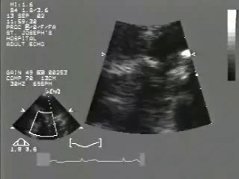
Diagnosis of aortic stenosis is suspected clinically and confirmed by echocardiography. Two-dimensional transthoracic echocardiography is used to identify a stenotic aortic valve and possible causes, to quantify LV hypertrophy and degree of systolic dysfunction, and to detect coexisting valvular heart disorders (aortic regurgitation, mitral valve disorders) and complications (eg, endocarditis). Doppler echocardiography is used to quantify degree of stenosis by measuring jet velocity, transvalvular systolic pressure gradient, and aortic valve area.
Severity of aortic stenosis is characterized echocardiographically as (1):
Mild: Peak aortic jet velocity 2.5 to 2.9 m/second, mean gradient 10 to 20 mm Hg, or valve area 1.5 to 2.0 cm2
Moderate: Peak aortic jet velocity 3 to 4 m/second, mean gradient 20 to 40 mm Hg, or valve area 1.0 to 1.5 cm2
Severe: Peak aortic jet velocity > 4 m/second, mean gradient > 40 mm Hg, or valve area < 1.0 cm2
Very severe: Peak aortic jet velocity > 5 m/second or mean gradient > 60 mm Hg
Clinical judgment and critical review of the data are used to resolve any discordance among these parameters (eg, moderate valve area but severe mean gradient). When LV function is normal, peak velocity is preferred as it is significantly more reproducible than valve area (2). Quantification of aortic valve stenosis is least accurate when LV volume or systolic function is reduced, or when systemic hypertension is present.
The gradient may be overestimated when aortic regurgitation is present. The gradient may under-represent severity when the stroke volume is low, eg, in patients with systemic hypertension or LV systolic dysfunction (low-gradient AS with reduced EF) or a small, hypertrophied LV (low-gradient AS with normal EF—see table Types of Severe Aortic Stenosis).
Sometimes LV systolic dysfunction results in low ventricular pressure that is inadequate to open nonstenotic valve leaflets, causing echocardiographic appearance of low valve area in the absence of stenosis (pseudo-stenosis). Differentiation of pseudo-stenosis from low-gradient AS can be aided by calculation of the ratio of outflow tract to aortic velocity (Doppler velocity index [DVI]). A DVI value < 0.25 suggests true severe stenosis.
Assessment of the degree of valve calcification by computed tomography (CT) can help determine the severity of AS. Severe AS is likely when the valvular calcium score is > 2000 Agatston units (AU) (men) and > 1300 AU (women). It is very likely when the calcium score is > 3000 AU(men) and > 1600 AU (women). Low-dose dobutamine stress echocardiography distinguishes low-gradient AS from pseudo-stenosis.
Types of Severe Aortic Stenosis
Severe AS type | Vmax | Valve area | LV EF | Other |
|---|---|---|---|---|
High gradient (asymptomatic or symptomatic) | ≥ 4.0 m/second | ≤ 1.0 cm2 | ≥ 50% | — |
Low-flow, low-gradient with reduced EF | < 4.0 m/second | ≤ 1.0 cm2 | < 50% | Distinguish from pseudo-stenosis with low-dose dobutamine echocardiography, valve area < 1.0 cm2, Vmax > 4.0 m/second |
Low-flow, low-gradient with normal EF (paradoxical low-flow AS) | < 4.0 m/second | ≤ 1.0 cm2 | ≥ 50% with small hypertrophied LV | Distinguish from non-severe AS by stroke volume index < 35 mL/m2 measured when patient is normotensive |
AS = aortic stenosis; DVI = Doppler velocity index, the ratio of LV outflow tract to aortic velocity; LV EF = left ventricular ejection fraction; Vmax = peak forward velocity across the aortic valve. | ||||
Data from Otto CM, Nishimura RA, Bonow RO, et al: 2020 ACC/AHA Guideline for the Management of Patients With Valvular Heart Disease: Executive Summary: A Report of the American College of Cardiology/American Heart Association Joint Committee on Clinical Practice Guidelines. Circulation 143(5):e35–e71, 2021. doi: 10.1161/CIR.0000000000000932 | ||||
Cardiac catheterization is performed prior to intervention to determine whether coronary artery disease (CAD) is the cause of angina and, occasionally, to resolve inconsistent clinical and echocardiographic estimates of AS severity.
An ECG and chest radiograph are obtained.
ECG typically shows changes of LV hypertrophy with or without an ischemic ST- and T-wave pattern.
Chest radiograph findings may include calcification of the aortic cusps (seen on the lateral projection or on fluoroscopy) and evidence of heart failure. Heart size may be normal or only mildly enlarged.
In asymptomatic patients with severe aortic stenosis, closely supervised exercise ECG testing is recommended in an attempt to elicit symptoms of angina, dyspnea, or hypotension—any of these symptoms, when due to the AS, is an indication for intervention. Failure to normally increase blood pressure and development of ST-segment depression are less predictive of adverse prognosis. Exercise testing is contraindicated in symptomatic patients.
Phonocardiographic Characteristics of Heart Murmurs


Diagnosis references
1. Otto CM, Nishimura RA, Bonow RO, et al: 2020 ACC/AHA Guideline for the Management of Patients With Valvular Heart Disease: Executive Summary: A Report of the American College of Cardiology/American Heart Association Joint Committee on Clinical Practice Guidelines. Circulation 143(5):e35–e71, 2021. doi: 10.1161/CIR.0000000000000932
2. Manna D, Eliasson M, Bech-Hanssen O, Lindow T. Reproducibility of Echocardiographic Measures of Aortic Stenosis Severity and its Impact on Grading of Severity. J Am Soc Echocardiogr 37(3):370–372.e2, 2024. doi:10.1016/j.echo.2023.11.006
Treatment of Aortic Stenosis
Sometimes aortic valve replacement
Nothing has been proved to slow the progression of aortic stenosis (1). Statins have been ineffective (2).
Medications that can cause hypotension (eg, nitrates) should be used cautiously, although nitroprusside has been used as a temporizing measure to reduce afterload in patients with decompensated heart failure in the hours before valve replacement. Medications that can cause hypotension (eg, nitrates) should be used cautiously, although nitroprusside has been used as a temporizing measure to reduce afterload in patients with decompensated heart failure in the hours before valve replacement.
Patients who develop heart failure but are too high risk for valve intervention benefit from cautious treatment with digoxin, diuretics, and angiotensin-converting enzyme (ACE) inhibitors.Patients who develop heart failure but are too high risk for valve intervention benefit from cautious treatment with digoxin, diuretics, and angiotensin-converting enzyme (ACE) inhibitors.
Indications for intervention
The benefits of intervention do not outweigh the risks until patients develop symptoms and/or meet certain echocardiographic criteria. Thus, patients should have periodic clinical evaluations, including echocardiography and sometimes exercise testing, to determine the optimal time for valve replacement (1). Valve replacement is recommended when aortic stenosis is severe and there is any one of the following:
Symptoms
Exercise testing causing symptoms or showing reduced effort tolerance or a fall in blood pressure ≥ 10 mm Hg below baseline)
LV EF < 50%
In addition, if surgical risk is low, then surgery may also be considered in asymptomatic patients with severe AS if there is any one of:
Very severe AS (aortic velocity > 5 m/second)
Brain (B-type) natriuretic peptide (BNP) is > 3 times normal
Severe pulmonary hypertension without other explanation
Decline in EF to < 60%
Severe valve calcification with rapid progression of stenosis (reduction in aortic valve area ≥ 0.3 m/second/year)
Evidence also supports surgical intervention in asymptomatic but severe AS.
When cardiac surgery is being performed for other reasons, concomitant aortic valve surgery is indicated regardless of symptoms if the AS is moderate or greater.
Choice of intervention
Transcatheter balloon valvuloplasty is used primarily in children and very young adults with congenital AS.
In older patients who are not candidates for surgery, balloon valvuloplasty has been used as a bridge to valve replacement, but this procedure has a high complication rate and provides only temporary relief. Transcatheter valve implantation, which can be performed with similar procedural risk even in sick, high-risk patients, is more common.
Surgical aortic valve replacement (SAVR) usually involves replacement with a mechanical or bioprosthetic valve. In younger patients, the patient’s own pulmonary valve can be used, with a bioprosthesis then used to replace the pulmonary valve (Ross procedure). The advantage of the Ross procedure rather than SAVR to replace the aortic valve only is increased durability of the procedure. The valve exposed to systemic pressure is the patient's own pulmonary valve, and this lasts longer than other replacement valves. The bioprosthesis in the pulmonary position tends to also last a relatively long time.
Transcatheter (percutaneous) aortic valve implantation (TAVI) (sometimes called transcatheter aortic valve replacement, or TAVR) is a less invasive method of aortic valve replacement.
TAVI valves are constructed differently than SAVR valves; TAVI valves are inserted through an artery in a collapsed configuration to the aortic valve site and are then deployed/expanded to replace the stenotic aortic valve.
The choice is between the up-front risk and morbidity of SAVR versus the unknown durability of TAVI valves beyond 10 years, although durability studies ranging up to 10 years suggest a structural valve degeneration rate < 10% within that time frame (3, 4, 5). Guidelines to inform shared decision-making suggest SAVR for patients < 65 years; TAVI is preferred for patients whose arteries are suitable for the transfemoral approach and who are > 80 years or who have a life expectancy of < 10 years. In patients aged 65 to 80 years whose arteries are suitable for transfemoral TAVI, the decision regarding SAVR or TAVI is determined based on individual patient characteristics (1). If the patient’s arteries are not suited to the transfemoral route, then SAVR is preferred as long as surgical risk is not prohibitive.
In patients with a life expectancy of < 1 year even with a successful procedure, intervention is not recommended.
Compared with SAVR, transfemoral TAVI has lower short-term mortality and less risk of stroke, major bleeding, and atrial fibrillation. TAVI also requires a shorter hospital stay, causes less pain, and permits more rapid return to activity; however, it also increases vascular complications and paravalvular regurgitation and increases the need for permanent pacemaker implantation and early repeat valve intervention. Indications for TAVI will expand as research includes younger and less symptomatic patients, although trials should be interpreted with caution due to potential flaws in methodology (6, 7).
Preoperative evaluation for CAD is indicated so that coronary artery bypass grafting (CABG) and valve replacement, if indicated, can be performed during the same procedure.
An aortic bioprosthetic valve requires anticoagulation for 3 to 6 months postoperatively, but a mechanical valve requires lifetime anticoagulation using warfarin. Long-term non-vitamin K oral anticoagulants or An aortic bioprosthetic valve requires anticoagulation for 3 to 6 months postoperatively, but a mechanical valve requires lifetime anticoagulation using warfarin. Long-term non-vitamin K oral anticoagulants orwarfarin can be considered in patients with a bioprosthetic valve and atrial fibrillation (see also Anticoagulation for patients with a prosthetic cardiac valve or native valve disease).
Treatment references
1. Otto CM, Nishimura RA, Bonow RO, et al: 2020 ACC/AHA Guideline for the Management of Patients With Valvular Heart Disease: Executive Summary: A Report of the American College of Cardiology/American Heart Association Joint Committee on Clinical Practice Guidelines. Circulation 143(5):e35–e71, 2021. doi: 10.1161/CIR.0000000000000932
2. Thiago L, Tsuji SR, Nyong J, et al: Statins for aortic valve stenosis. Cochrane Database Syst Rev 9(9):CD009571, 2016. doi:10.1002/14651858.CD009571.pub2
3. Ali N, Hildick-Smith D, Parker J, et al: Long-term durability of self-expanding and balloon-expandable transcatheter aortic valve prostheses: UK TAVI registry. Catheter Cardiovasc Interv 101(5):932–942, 2023. doi:10.1002/ccd.30627
4. Blackman DJ, Saraf S, MacCarthy PA, et al: Long-Term Durability of Transcatheter Aortic Valve Prostheses. J Am Coll Cardiol 73(5):537–545, 2019. doi:10.1016/j.jacc.2018.10.078
5. Carrabba N, Migliorini A, Fumagalli C, et al: Long-Term Durability of Transcatheter Aortic Valve Implantation With Self-Expandable Valve System (from a Real-World Registry). Am J Cardiol 143:104–110, 2021. doi:10.1016/j.amjcard.2020.12.032
6. Généreux P, Schwartz A, Oldemeyer JB, et al: Transcatheter Aortic-Valve Replacement for Asymptomatic Severe Aortic Stenosis. N Engl J Med 392(3):217–227, 2025. doi:10.1056/NEJMoa2405880
7. Rajkumar CA, Nijjer SS, Cole GD, Al-Lamee R, Francis DP: 'Faith Healing' and 'Subtraction Anxiety' in Unblinded Trials of Procedures: Lessons from DEFER and FAME-2 for End Points in the ISCHEMIA Trial [published correction appears in Circ Cardiovasc Qual Outcomes. 2018 Apr;11(4):e000038. doi: 10.1161/HCQ.0000000000000038.. Title corrected]. Circ Cardiovasc Qual Outcomes 11(3):e004665., 2018 doi:10.1161/CIRCOUTCOMES.118.004665
Prognosis for Aortic Stenosis
Aortic stenosis progresses faster as severity increases, but the wide variability in progression rates requires regular surveillance, particularly in sedentary older patients. In such patients, flow may become significantly compromised without triggering symptoms.
Asymptomatic patients with severe AS and normal systolic function should be reevaluated every 6 months because up to 11% may have a fall in LVEF after one year(1). The risk of surgery outweighs the survival benefit in asymptomatic patients, but with the onset of symptoms the mean survival drops to 3 years (2), and prompt valve replacement is indicated to relieve symptoms and improve survival. Risk of surgery increases for patients who require simultaneous CABG and for those with depressed systolic LV function.
In patients with severe AS, approximately 13% of deaths occur suddenly (3), so patients with severe AS should be advised to limit physical exertion.
Prognosis references
1. Minamino-Muta E, Kato T, Morimoto T, et al. Decline in Left Ventricular Ejection Fraction During Follow-Up in Patients With Severe Aortic Stenosis. JACC Cardiovasc Interv 2019;12(24):2499-2511. doi:10.1016/j.jcin.2019.09.015
2. Carabello BA. Introduction to aortic stenosis. Circ Res 2013;113(2):179-185. doi:10.1161/CIRCRESAHA.113.300156
3. Minamino-Muta E, Kato T, Morimoto T, et al. Causes of Death in Patients with Severe Aortic Stenosis: An Observational study. Sci Rep 2017;7(1):14723. Published 2017 Nov 7. doi:10.1038/s41598-017-15316-6
Key Points
The most common cause of aortic stenosis (AS) is bicuspid aortic valve in patients < 70 years and aortic sclerosis in older patients.
Untreated AS ultimately results in exertional syncope, angina, and dyspnea; sudden death may occur.
Typical heart sounds are a crescendo-decrescendo ejection murmur that increases with maneuvers that increase left ventricular (LV) volume and contractility (eg, leg-raising, squatting, Valsalva release) and decreases with maneuvers that decrease LV volume (Valsalva maneuver) or increase afterload (isometric handgrip).
Nitrates may cause dangerous hypotension and should be used with caution for angina in patients with AS.
Exercise testing is useful for risk stratification and determining the need for surgery for patients without clear symptoms.
Valve replacement is indicated once symptoms begin or LV dysfunction occurs.
Surgical or transcatheter aortic valve replacement are options for many patients, but long-term safety data are still accumulating for transcatheter aortic valve replacement (especially in low-risk patients).


