The spinal cord is a long, fragile tubelike structure that begins at the end of the brain stem and continues down almost to the bottom of the spine. The spinal cord consists of bundles of nerve axons forming pathways that carry incoming and outgoing messages between the brain and the rest of the body. The spinal cord contains nerve cell circuits that control coordinated movements such as walking and swimming, as well as urinating. It is also the center for reflexes, such as the knee jerk reflex (see figure Reflex Arc: A No-Brainer).

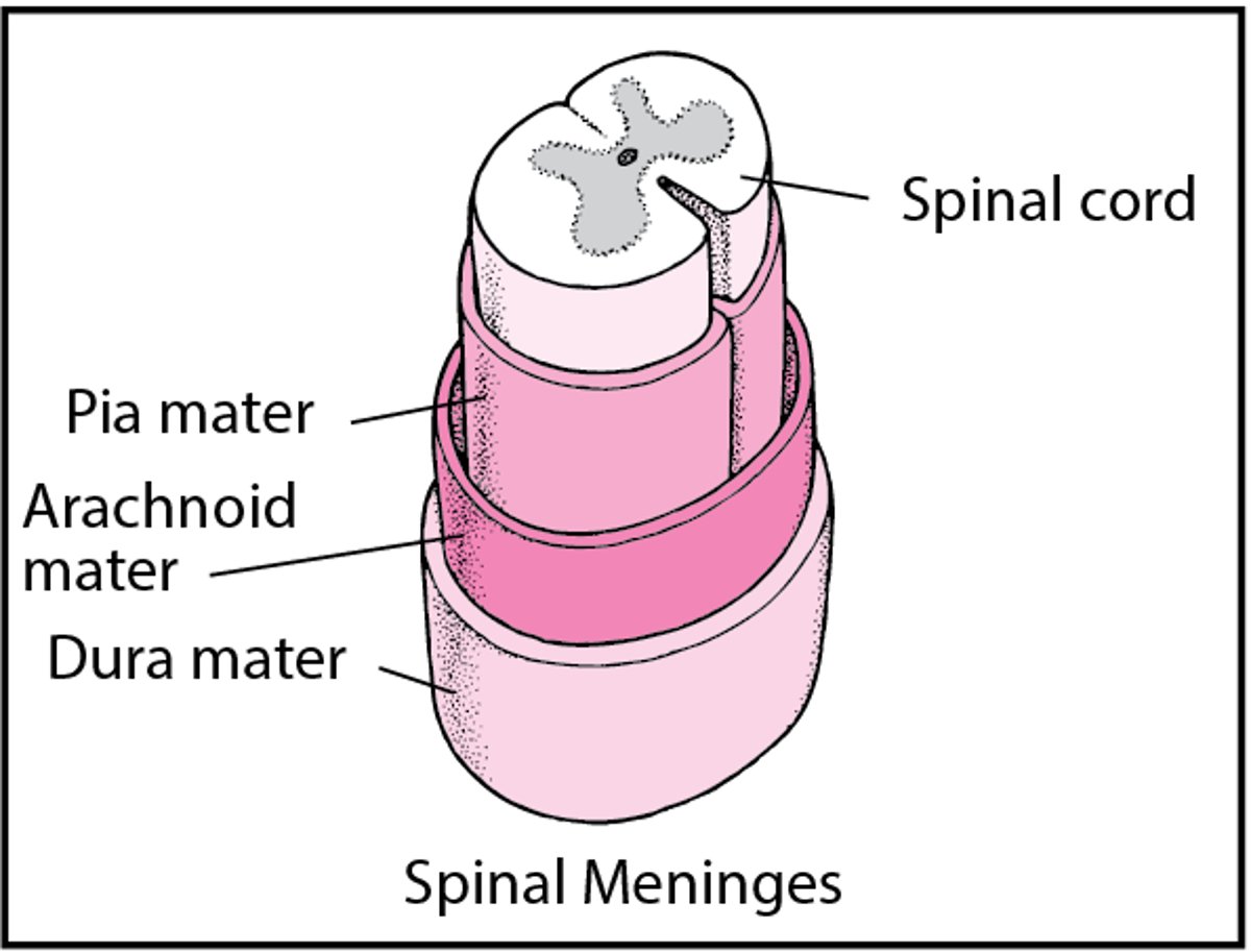
Like the brain, the spinal cord is covered by 3 layers of tissue (meninges). The spinal cord and meninges are contained in the spinal canal, which runs through the center of the spine. In most adults, the spine is composed of 33 individual back bones (vertebrae). Just as the skull protects the brain, vertebrae protect the spinal cord. The vertebrae are separated by disks made of cartilage, which act as cushions, reducing the forces on the spine generated by movements such as walking and jumping. The vertebrae and disks of cartilage extend the length of the spine and together form the vertebral (spinal) column.
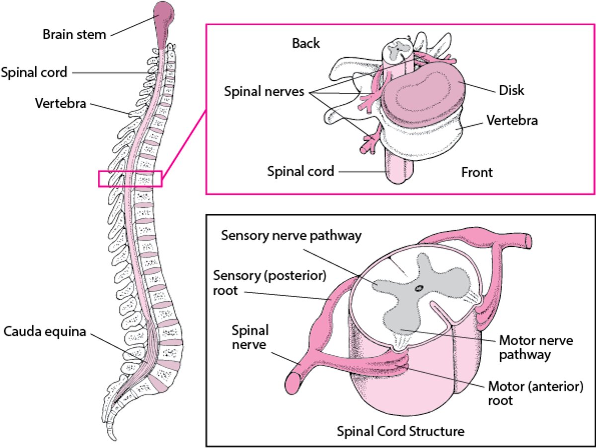
How the Spine Is Organized
A column of bones called vertebrae make up the spine (spinal column). The vertebrae protect the spinal cord, a long, fragile structure contained in the spinal canal, which runs through the center of the spine. Between the vertebrae are disks composed of cartilage, which help cushion the spine and give it some flexibility. Like the brain, the spinal cord is covered by 3 layers of tissue (meninges). | |
Spinal nerves: 31 pairs of spinal nerves emerge from the spinal cord between the vertebrae. Each nerve emerges in 2 short branches (roots):
The motor roots carry commands from the brain and spinal cord primarily to skeletal muscles to control movement. The sensory roots carry sensory information (about pain, temperature, vibration, limb position, and light touch) to the brain from other parts of the body. Cauda equina: The spinal cord ends about three-fourths of the way down the spine, but a bundle of nerves extends beyond the cord. This bundle is called the cauda equina because it resembles a horse’s tail. The cauda equina carries nerve impulses, both motor and sensory, to and from the legs. | |


Like the brain, the spinal cord consists of gray and white matter.
The gray matter forms a butterfly-shaped center in the cord. The front wings (called anterior or ventral horns) contain motor nerve cells (neurons) which transmit information from the brain or spinal cord to muscles, stimulating movement. The back part of the butterfly wing (called posterior or dorsal horns) contains sensory nerve cells, which transmit sensory information from other parts of the body through the spinal cord to the brain.
The surrounding white matter contains columns of nerve fibers (axon bundles) that carry sensory information to the brain from the rest of the body (ascending tracts) and columns that carry motor impulses from the brain to the muscles (descending tracts).