- Introduction to the Neurologic Examination
- How to Assess Mental Status
- How to Assess the Cranial Nerves
- How to Assess the Motor System
- How to Assess Muscle Strength
- How to Assess Gait, Stance, and Coordination
- How to Assess Sensation
- How to Assess Reflexes
- How to Assess the Autonomic Nervous System
- Cerebrovascular Examination
Topic Resources
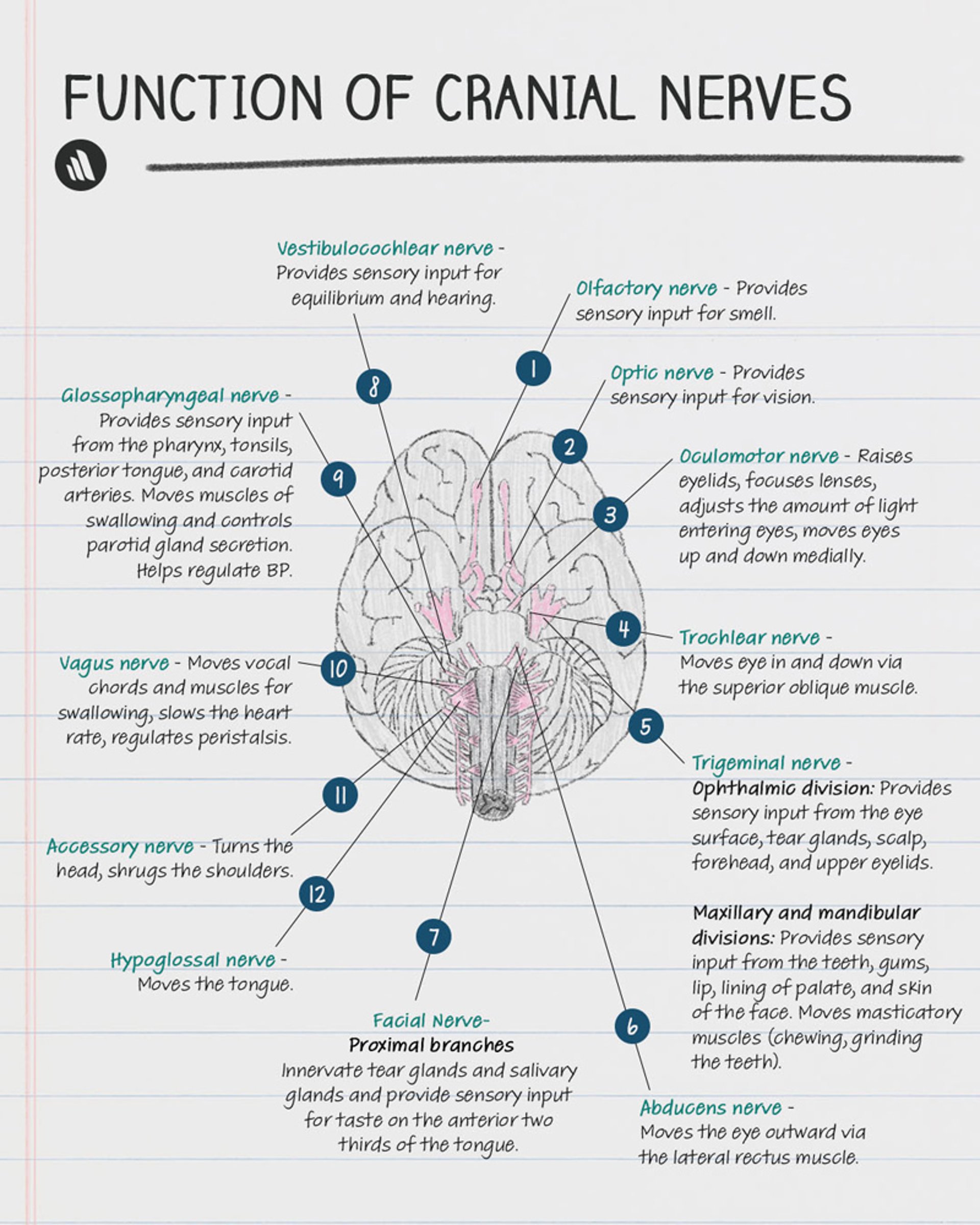
The cranial nerves originate in the brain stem. Abnormalities in their function suggest pathology in specific parts of the brain stem or along the cranial nerve's path outside the brain stem. For example, unilateral leg weakness with upper motor signs may be due to pathology anywhere between the cerebral cortex and the lumbar spine. However, the presence of an abnormal cranial nerve sign strongly suggests that the observed weakness results from a problem in the brain stem. Specific combinations of cranial nerve signs may suggest pathology at specific locations around the base of the skull.
Copyright © 2023 Merck & Co., Inc., Rahway, NJ, USA and its affiliates. All rights reserved.
(See also Neuro-ophthalmologic and Cranial Nerve Disorders and Introduction to the Neurologic Examination.)
1st Cranial nerve
Smell, a function of the 1st (olfactory) cranial nerve, is usually evaluated only after head trauma or when lesions of the anterior fossa (eg, meningioma) are suspected or patients report abnormal smell or taste.
The patient is asked to identify odors (eg, soap, coffee, cloves) presented to each nostril while the other nostril is occluded. Alcohol, ammonia, and other irritants, which test the nociceptive receptors of the 5th (trigeminal) cranial nerve, are used only when malingering is suspected.
2nd Cranial nerve
For the 2nd (optic) cranial nerve, visual acuity is tested using a Snellen chart for distance vision or a handheld chart for near vision; each eye is assessed individually, with the other eye covered.
Color perception is tested using standard pseudoisochromatic Ishihara or Hardy-Rand-Ritter plates that have numbers or figures embedded in a field of specifically colored dots.
Visual fields are tested by directed confrontation in all 4 visual quadrants. Direct and consensual pupillary responses are tested. Funduscopic examination is also done.
3rd, 4th, and 6th Cranial nerves
For the 3rd (ocolomotor), 4th (trochlear), and 6th (abducens) cranial nerves, eyes are observed for symmetry of movement, globe position, asymmetry or droop of the eyelids (ptosis), and twitches or flutters of globes or lids. Extraocular movements controlled by these nerves are tested by asking the patient to follow a moving target (eg, examiner’s finger, penlight) to all 4 quadrants (including across the midline) and toward the tip of the nose; this test can detect nystagmus and palsies of ocular muscles. Brief fine amplitude nystagmus at end-lateral gaze is normal.
Anisocoria or differences in pupillary size should be noted in a dimly lit room. The pupillary light response is tested for symmetry and briskness.
5th Cranial nerve
For the 5th (trigeminal) nerve, the 3 sensory divisions (ophthalmic, maxillary, mandibular) are evaluated by using a pinprick to test facial sensation and by brushing a wisp of cotton against the lower or lateral cornea to evaluate the corneal reflex. If facial sensation is lost, the angle of the jaw should be examined; sparing of this area (innervated by spinal root C2) suggests a trigeminal deficit. A weak blink due to facial weakness (eg, 7th cranial nerve paralysis) should be distinguished from depressed or absent corneal sensation, which is common in contact lens wearers. A patient with facial weakness feels the cotton wisp normally on both sides, even though blink is decreased.
Trigeminal motor function is tested by palpating the masseter muscles while the patient clenches the teeth and by asking the patient to open the mouth against resistance. If a pterygoid muscle is weak, the jaw deviates to that side when the mouth is opened.
7th Cranial nerve
The 7th (facial) cranial nerve is evaluated by checking for hemifacial weakness. Asymmetry of facial movements is often more obvious during spontaneous conversation, especially when the patient smiles or, if obtunded, grimaces at a noxious stimulus; on the weakened side, the nasolabial fold is depressed and the palpebral fissure is widened. If the patient has only lower facial weakness (ie, furrowing of the forehead and eye closure are preserved), etiology of 7th nerve weakness is central rather than peripheral.
Taste in the anterior two thirds of the tongue can be tested with sweet, sour, salty, and bitter solutions applied with a cotton swab first on one side of the tongue, then on the other.
Hyperacusis, indicating weakness of the stapedius muscle, may be detected with a vibrating tuning fork held next to the ear.
8th Cranial nerve
Because the 8th (vestibulocochlear, acoustic, auditory) cranial nerve carries auditory and vestibular input, evaluation involves
Vestibular function tests
Hearing is first tested in each ear by whispering something while occluding the opposite ear. Any suspected loss should prompt formal audiologic testing to confirm findings and help differentiate conductive hearing loss from sensorineural hearing loss. The Weber and Rinne tests may be done at the bedside to attempt to differentiate the two, but they are difficult to do effectively except in specialized settings.
Vestibular function can be evaluated by testing for nystagmus. The presence and characteristics (eg, direction, duration, triggers) of nystagmus help identify vestibular disorders and sometimes differentiate central from peripheral vertigo. Vestibular nystagmus has 2 components:
A slow component caused by vestibular input
A quick, corrective component that causes movement in the opposite direction (called beating)
The direction of the nystagmus is defined by the direction of the quick component because it is easier to see. Nystagmus may be rotary, vertical, or horizontal and may occur spontaneously, with gaze, or with head motion.
When trying to differentiate central from peripheral causes of vertigo, the following guidelines are reliable and should be considered at the onset:
There are no central causes of unilateral hearing loss because peripheral sensory input from the 2 ears is combined virtually instantaneously as the peripheral nerves enter the pons.
There are no peripheral causes of CNS signs. If a CNS sign (eg, cerebellar ataxia) appears at the same time as the vertigo, the localization is virtually certain to be central.
Evaluation of vertigo using nystagmus testing is particularly useful in the following situations:
When patients are having vertigo during the examination
When patients have acute vestibular syndrome
When patients have episodic, positional vertigo
If patients have acute vertigo during the examination, nystagmus is usually apparent during inspection. However, visual fixation can suppress nystagmus. In such cases, the patient is asked to wear +30 diopter or Frenzel lenses to prevent visual fixation so that nystagmus, if present, can be observed. Clues that help differentiate central from peripheral vertigo in these patients include the following:
If nystagmus is absent with visual fixation but present with Frenzel lenses, it is probably peripheral.
If nystagmus changes direction (eg, from one side to the other when, for example, when the direction of gaze changes), it is probably central. However, absence of this finding does not exclude central causes.
If nystagmus is peripheral, the eyes beat away from the dysfunctional side.
When evaluating patients with acute vestibular syndrome (rapid onset of severe vertigo, nausea and vomiting, spontaneous nystagmus, and postural instability), the most important maneuver to help differentiate central vertigo from peripheral vertigo is the head thrust maneuver. With the patient sitting, the examiner holds the patient's head and asks the patient to focus on an object, such as the examiner's nose. The examiner then suddenly and rapidly turns the patient's head about 20° to the right or left. Normally, the eyes stay focused on the object (via the vestibular ocular reflex). Other findings are interpreted as follows:
If the eyes temporarily move away from the object and then a frontal corrective saccade returns the eyes to the object, nystagmus is probably peripheral (eg, vestibular neuronitis). The vestibular apparatus on one side is dysfunctional. The faster the head is turned, the more obvious is the corrective saccade.
If the eyes stay focused on the object and there is no need for a corrective saccade, nystagmus is probably central (eg, cerebellar stroke).
When vertigo is episodic and provoked by positional change, the Dix-Hallpike (or Barany) maneuver is done to test for obstruction of the posterior semicircular canal with displaced otoconial crystals (ie, for benign paroxysmal positional vertigo [BPPV]). In this maneuver, the patient sits upright on the examining table. The patient is rapidly lowered backward to a supine position with the head extended 45° below the horizontal plane (over the edge of the examining table) and rotated 45° to one side (eg, to the right side). Direction and duration of nystagmus and development of vertigo are noted. The patient is returned to an upright position, and the maneuver is repeated with rotation to the other side. Nystagmus secondary to BPPV has the following nearly pathognomic characteristics:
A latency period of 5 to 10 seconds
Usually, vertical (upward-beating) nystagmus when the eyes are turned away from the affected ear and rotary nystagmus when the eyes are turned toward the affected ear
Nystagmus that fatigues when the Dix-Hallpike maneuver is repeated
In contrast, positional vertigo and nystagmus related to CNS dysfunction have no latency period and do not fatigue.
The Epley canalith repositioning maneuver (see figure Epley Maneuver) can be done for both sides to help confirm the diagnosis of BPPV. If the patient has BPPV, there is a high probability (up to 90%) that the symptoms will disappear after the Epley maneuver, and results of a repeat Dix-Hallpike maneuver will then be negative.
9th and 10th Cranial nerves
The 9th (glossopharyngeal) and 10th (vagus) cranial nerves are usually evaluated together. Whether the palate elevates symmetrically when the patient says "ah" is noted. If one side is paretic, the uvula is lifted away from the paretic side. A tongue blade can be used to touch one side of the posterior pharynx, then the other, and symmetry of the gag reflex is observed; bilateral absence of the gag reflex is common among healthy people and may not be significant.
In an unresponsive, intubated patient, suctioning the endotracheal tube normally triggers coughing.
If hoarseness is noted, the vocal cords are inspected. Isolated hoarseness (with normal gag and palatal elevation) should prompt a search for lesions (eg, mediastinal lymphoma, aortic aneurysm) compressing the recurrent laryngeal nerve.
11th Cranial nerve
The 11th (spinal accessory) cranial nerve is evaluated by testing the muscles it supplies:
For the sternocleidomastoid, the patient is asked to turn the head against resistance supplied by the examiner’s hand while the examiner palpates the active muscle (opposite the turned head).
For the upper trapezius, the patient is asked to elevate the shoulders against resistance supplied by the examiner.
12th Cranial nerve
The 12th (hypoglossal) cranial nerve is evaluated by asking the patient to extend the tongue and inspecting it for atrophy, fasciculations, and weakness (deviation is toward the side of a lesion).


